Zika Virus and Birth Defects: How Strong is the Correlation?
BACKGROUND AND PURPOSE:
Risk of congenital neurologic defects related to Zika virus (ZIKV) infection has ranged from 6 to 42% in contrasting reports
The magnitude of risk is still unclear
Hoen et al. (N Eng j Med, 2018) aimed to assess risk among pregnant women with symptomatic ZIKV infection in French territories (Guadeloupe, Martinique, Guyane) in the Americas
METHODS:
Prospective cohort study
Participants: Pregnant women with
Symptomatic ZIKV infection, confirmed by polymerase-chain-reaction (PCR) assay (blood, urine or both)
Symptomatic: At least one of the following consistent with acute ZIKV infection
Pruritic rash, fever, conjunctival hyperemia, arthralgia, or myalgia
Microcephaly: Head circumference >2 SD below the mean for sex and gestational age
Severe microcephaly: Head circumference >3 SD below the mean
The data was collected up until the last delivery in the cohort
RESULTS:
Data was collected from 555 fetuses and infants among the 546 pregnancies that were included in the analysis
28 (5.0%) were not carried to term
527 were born alive
Neurologic and ocular defects possibly associated with ZIKV infection were seen in 7% of fetuses and infants (95% CI, 5.0 to 9.5); of these
10 were not carried to term because of termination of pregnancy for medical reasons
1 was stillborn
28 were live-born
Microcephaly was detected in
5.8% fetuses and infants and of those
1.6% had severe microcephaly
Neurologic and ocular defects were more common when ZIKV infection occurred during the first trimester (P=0.001)
First trimester 12.7%
Second trimester 3.6%
Third trimester 5.3%
CONCLUSION:
Defects associated with ZIKV were found in 7% of fetuses and infants from mothers with ZIKV
Overall risk of evident birth defects included in the current definition of the congenital Zika syndrome was 3.1% (see ‘Related ObG Topics’ below)
There was an increased risk of birth defects when infection occurs earlier in pregnancy
Zika virus disease is a nationally notifiable condition:
Healthcare providers should report suspected Zika virus disease cases to their state, local, or territorial health department, who will report to the CDC.
WHAT IS IT?
The Zika virus (ZIKV) was first discovered in 1947, but has only become a global public health threat in the last decade with marked geographic spread in the last 5 years
The Zika virus is named after the Zika forest in Uganda where it was first found, is a Flavivirus and is related to similar viruses such as dengue, West Nile and Japanese encephalitis
The infectious viral particle (virion) is primarily composed of a single strand of RNA which is released into the infected cell’s cytoplasm, overtakes the infected cell’s genetic and cellular machinery, leading to replication and release of additional Zika virus
ZIKV, typical for a Flavirus, is predominantly spread via mosquito vectors, however transmission via blood transfusion and sexual contact can occur
KEY POINTS:
Clinical symptoms appear 3 to 14 days after a mosquito bite and should resolve within 2 to 7 days
Most people will be asymptomatic
20% will have typical viral signs and symptoms, usually of a mild nature
Fever
Maculopaular rash
Conjunctivitis
Arthralgias
Headache
Management is that of typical viral illness and includes:
Rest
Antipyretics
Note: current drug labels state that NSAIDs should not be used by pregnant women in their third trimester of pregnancy because of the risk of premature closure of the ductus arteriosus in the fetus
Nutrition and adequate fluids
Monitor for signs and symptoms of severe infection such as coagulopathies and organ damage – ICU care is rare but any concern should be escalated
Guillain-Barré syndrome, an autoimmune neurological disorder, has been reported and while uncommon should generate a referral for diagnosis and management
Microcephaly means “small head”, and can be used interchangeably in some instances with microencephaly, “small brain”
Consider abnormal development (primary microcephaly) if detected prior to 32 weeks gestation, as opposed to a degenerative condition in which normal head size becomes smaller (secondary microcephaly)
SYNOPSIS:
Microcephaly has an incidence of 2 to 12 in 10,000 births in the USA and can be diagnosed prenatally via ultrasound (in second or early third trimester) or postnatally via measurement of head circumference (HC). Microcephaly has been linked to developmental delay, seizures, as well as feeding, vision and hearing problems. Prognosis depends on the severity of the microcephaly and whether it is associated with other anomalies.
KEY POINTS:
Definitions
SMFM – Fetal Microcephaly
HC ≥ 3 SD below the mean for gestational age is the recommended definition
HC ≥ 5 SD below the mean is a certain diagnosis
CDC – Postnatal Congenital Microcephaly
Definite Congenital Microcephaly
HC at birth < 3rd percentile for gestational age and sex
If HC not available at birth, HC < 3rd percentile for age and sex within the first 2 weeks of life
For Stillbirths and elective terminations, CDC definition based on HC at delivery < 3rd percentile for gestational age and sex
Possible Congenital Microcephaly
For Live Births: If earlier HC is not available, HC < 3rd percentile for age and sex beyond 6 weeks of life
For All Pregnancy Outcomes: Microcephaly diagnosed or suspected on prenatal ultrasound in the absence of available postnatal HC measurements
American Academy of Neurology and Child Neurology Society
At birth
HC >2 SD below the mean for age and gender
Severe would be considered >3 SD below the mean
Management
If fetal HC by ultrasound is > 2 SD below the mean for gestational age, a careful evaluation of the fetal intracranial anatomy is indicated
If the intracranial anatomy is normal, consider follow-up in 3-4 weeks
When evaluating the finding of fetal microcephaly, assess for the following
Alcohol consumption, exposure to certain medications and/or smoking during pregnancy
Genetic conditions, including aneuploidy or single gene disorders
Many ultrasound packages report HC percentiles and not standard deviations (SD) and often the lowest reported HC measurement is <5th percentile
In such cases, SMFM suggests using head circumference (in millimeters) to determine SD below the mean
SMFM provides a table (see ‘Learn More – Primary Sources’ below) to determine the number of SD below the mean as a function of gestational age
Role of MRI: The SOGC Guideline states (III-A evidence)
Once fetal microcephaly is identified by ultrasound, fetal magnetic resonance imaging, when available and if potential findings are likely to alter pregnancy management, may be considered.
Fetal magnetic resonance imaging images should be reviewed by a radiologist with expertise in fetal magnetic resonance imaging.
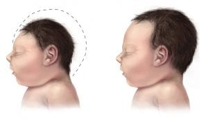
Consider referral to a Fetal Maternal Medicine Specialist
Consider referral to Genetic Counseling, especially if microcephaly is associated with other findings
Consider referral to Pediatric Neurologist for review of short and long term potential outcomes for the newborn and child with microcephaly
Image credit: Centers for Disease Control and Prevention, National Center on Birth Defects and Developmental Disabilities
OBG Project CME requires a modern web browser (Internet Explorer 10+, Mozilla Firefox, Apple Safari, Google Chrome, Microsoft Edge). Certain educational activities may require additional software to view multimedia, presentation, or printable versions of their content. These activities will be marked as such and will provide links to the required software. That software may be: Adobe Flash, Apple QuickTime, Adobe Acrobat, Microsoft PowerPoint, Windows Media Player, or Real Networks Real One Player.
Disclosure of Unlabeled Use
This educational activity may contain discussion of published and/or investigational uses of agents that are not indicated by the FDA. The planners of this activity do not recommend the use of any agent outside of the labeled indications.
The opinions expressed in the educational activity are those of the faculty and do not necessarily represent the views of the planners. Please refer to the official prescribing information for each product for discussion of approved indications, contraindications, and warnings.
Disclaimer
Participants have an implied responsibility to use the newly acquired information to enhance patient outcomes and their own professional development. The information
presented in this activity is not meant to serve as a guideline for patient management. Any procedures, medications, or other courses of diagnosis or treatment discussed or suggested in this activity should not be used by clinicians without evaluation of their patient’s conditions and possible contraindications and/or dangers in use, review of any applicable manufacturer’s product information, and comparison with recommendations of other authorities.
Jointly provided by
NOT ENOUGH CME HOURS
It appears you don't have enough CME Hours to take this Post-Test. Feel free to buy additional CME hours or upgrade your current CME subscription plan
You are now leaving the ObG website and on your way to PRIORITY at UCSF, an independent website. Therefore, we are not responsible for the content or availability of this site