For Physicians. By Physicians.™

ObGFirst: Get guideline notifications, fast. First month free!Click here
Practical obstetrics info for your women's healthcare practice

Gastroschisis – Detection and Implications

WHAT IS IT? 

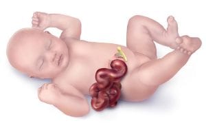
  • Gastroschisis is the presence of intestinal loops in the amniotic cavity due to failure of abdominal body wall closure, typically to the right of the umbilicus
  • There is no membrane covering the protruding organs, therefore organs are exposed to amniotic fluid and can be damaged
  • Incidence 1/1800 births per year in USA
  • Chromosomal abnormalities are not typically associated, unlike omphalocele
    • One international study reported chromosomal anomalies in 1.2% (total 3,322 cases) with the most frequent being
      • Trisomy 18 (15 cases) | Trisomy 13 (14 cases) | Sex chromosomes anomalies (4 cases) | Trisomy 21 (2 cases)

KEY CLINICAL POINTS:

  • Cause of gastroschisis unknown in most cases
  • Associations have been demonstrated with maternal history of
    • Young age and thin body type
    • Primipara or primigravida females
    • Cigarette smoking in pregnancy
    • Poor maternal diet
    • Low socioeconomic status
    • Maternal use of certain medications (phenylpropanolamine and pseudoephedrine decongestants, salicylates, ibuprofen, acetaminophen)
  • Prenatal screening and detection is available
    • Maternal serum AFP (MSAFP) is elevated
    • Amniotic fluid AFP (AFAFP) is elevated
    • Possible polyhydramnios detected on prenatal ultrasound
  • If gastroschisis is detected on ultrasonography 
    • ACOG guidance recommends offering invasive testing using microarray in the setting of fetal structural anomalies seen on prenatal ultrasound
    • Fetal echocardiography referral
    • Ensure detailed anatomy ultrasound that includes visualization of all limbs, to rule out potential syndrome associated with body-stalk disruption
      • Note abdominal opening to the right of the umbilicus
    • Minimal recurrence risk 2.5%
  • Refer to Pediatric surgical specialist to discuss possible surgical correction options, outcomes and long term implications
  • Develop labor and delivery plan for those who will continue the pregnancy given its high risk nature, and coordinate with NICU and pediatric team as needed
  • Data suggests better outcomes at perinatal centers that also include surgical teams capable of definitive operative management and NICU availability
Gastroschisis

Image credit: Centers for Disease Control and Prevention, National Center on Birth Defects and Developmental Disabilities


Want to hear about the latest clinical summaries via ObG Insider?

Get Your Free e-Newsletter »


Learn More – Primary Sources:

CDC: Facts about Gastroschisis

Gastroschisis and associated defects: an international study

Review of Abdominal Wall Defects

Prenatal Risk Factors and Outcomes in Gastroschisis: A Meta-Analysis

Gastroschisis: antenatal sonographic predictors of adverse neonatal outcome

Influence of location of delivery on outcome in neonates with gastroschisis

Locate a Maternal Fetal Medicine Specialist:

Maternal-Fetal Medicine Specialist Locator-SMFM

Atrial Septal Defect (ASD) – Summary and Implications

WHAT IS IT? 

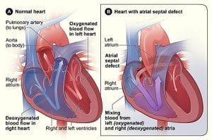
  • Atrial septal defects can be generally classified into 3 subtypes:
    • Ostium secundum ASD: defect of the atrial septum that involved the region of the fossa ovalis
      • More common in females (65-75%)
      • While endocardial cushion defects are more commonly associated with Down syndrome, secundum ASDs have also been associated with Down syndrome
    • Ostium primum ASD: actually an atrioventricular (AV) septal defect, sometimes referred to as an AV canal defect or endocardial cushion defect
      • Associated with Down syndrome (15 to 20%) but may also be associated with other chromosomal abnormalities such as DiGeorge syndrome (22q11 deletion syndrome)
    • Sinus venosus ASD: occurs at the junction of the right atrium and superior vena cava and is almost always associated with partial anomalous pulmonary venous return
  • May be associated with arrhythmias
  • Other uncommon subtypes do exist
Atrial Septal Defect

Image credit – National Heart Lung and Blood Institute (NIH)

CAUSE:

ASD is typically sporadic, however may occur as an isolated finding in families or as part of various genetic syndromes including but not limited to Down syndrome, DiGeorge syndrome and Holt-Oram syndrome which involves variable radial ray anomalies.

KEY POINTS:

  • Can range from small isolated finding that will close on its own, to complex congenital heart defect (CHD) requiring surgical repair and interdisciplinary team management
  • Consider genetic consultation to identify potential cause as may influence management
    • ACOG guidance recommends offering invasive testing using microarray in the setting of fetal structural anomalies seen on prenatal ultrasound

Learn More – Primary Sources: 

AHA Circulation Journal -Atrial Septal Defects in the Adult

CDC: Facts about Atrial Septal Defect

ACOG Practice Bulletin No. 162: Prenatal Diagnostic Testing for Genetic Disorders

Locate a Genetic Counselor or Genetics services:

Genetic Services Locator-ACMG

Genetic Services Locator-NSGC

Genetic Services Locator-CAGC