Evidence-Based. Clearly Delivered.
Evidence-Based. Clearly Delivered.

A microarray describes a newer technology that can identify small duplications or deletions of genetic material that previously could not be identified using conventional karyotyping alone. It has become a critical tool to help identify submicroscopic chromosomal deletions/duplications that underlie clinically significant syndromes in the prenatal period and throughout the lifespan.
What is a Deletion?
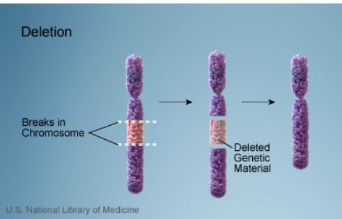
Credit: US National Library of Medicine
What is a Microdeletion?
What is a CNV (copy number variant)?
CGH (Comparative genome hybridization)
SNP array
Additional capabilities of SNP array compared to CGH
SMFM Consult Series 41: The use of chromosomal microarray for prenatal diagnosis
Please log in to ObGFirst to access this page
OBG Project CME requires a modern web browser (Internet Explorer 10+, Mozilla Firefox, Apple Safari, Google Chrome, Microsoft Edge). Certain educational activities may require additional software to view multimedia, presentation, or printable versions of their content. These activities will be marked as such and will provide links to the required software. That software may be: Adobe Flash, Apple QuickTime, Adobe Acrobat, Microsoft PowerPoint, Windows Media Player, or Real Networks Real One Player.
This educational activity may contain discussion of published and/or investigational uses of agents that are not indicated by the FDA. The planners of this activity do not recommend the use of any agent outside of the labeled indications.
The opinions expressed in the educational activity are those of the faculty and do not necessarily represent the views of the planners. Please refer to the official prescribing information for each product for discussion of approved indications, contraindications, and warnings.
Participants have an implied responsibility to use the newly acquired information to enhance patient outcomes and their own professional development. The information
presented in this activity is not meant to serve as a guideline for patient management. Any procedures, medications, or other courses of diagnosis or treatment discussed or suggested in this activity should not be used by clinicians without evaluation of their patient’s conditions and possible contraindications and/or dangers in use, review of any applicable manufacturer’s product information, and comparison with recommendations of other authorities.
Jointly provided by