HPV Vaccine Recommendations Including Guidance for Ages 27 to 45
SUMMARY:
Previously, US guidelines called for two doses when vaccination began before age 15, and three doses for those starting at age 15 or older or for individuals who were immunocompromised. The Department of Health and Human Services (HHS) supports a one‑dose recommendation based on international alignment with other high‑income countries and emerging scientific evidence indicating that one dose provides protection comparable to two doses. This directive has been published by the CDC. However, it is important to note that the US does not yet have a single‑dose HPV vaccine licensed for use. Furthermore, ACOG and AAP, based on current evidence, do not support this change in dosing.
HHS/CDC Recommendations
- Routine vaccination
- Age 11 to 12 years
- Can be started at age 9 years
- Catch-up vaccination
- Age 13 to 26 years
- If not adequately vaccinated
- Shared clinical decision-making
- Some adults ages 27 to 45 years
- If not adequately vaccinated
Dosing Schedule
- HHS/CDC
- Change to 1-dose regimen for children and adolescents
- AAP and ACOG
- <15 years: 2 doses spaced 6 to 12 months apart
- ≥15 years: 3-dose schedule
- Initial dose | Second dose at 1 to 2 months | Third dose at 6 months
ACOG HPV vaccine recommendations
- Routine HPV vaccination is recommended for females and males
- Target age is 11 to 12 years but can be given through age 26
- Can be given from age of 9
- Do not test for HPV DNA prior to vaccination
- Vaccinate even if patient was tested and is HPV DNA positive
- If not vaccinated between 11 to 12 years
- Vaccinate between 13 to 26 years (catch up period)
- Offer regardless of sexual activity, prior exposure to HPV, or sexual orientation
- Women 27 to 45 years and previously unvaccinated
- Use shared clinical decision making
- ACOG “does not recommend that an individual who received the quadrivalent HPV vaccine be revaccinated with 9-valent HPV vaccine, including those aged 27 to 45 years who previously completed some, but not all, of the vaccine series when they were younger”
- Pregnancy
- HPV vaccine is not recommended during pregnancy
- Pregnancy testing prior to HPV vaccination not recommended
- If vaccination schedule is interrupted by pregnancy, resume postpartum with the next dose
- HPV vaccine can and should be given to breastfeeding women ≤26 who have not been vaccinated
- The CDC encourages pregnant patients and healthcare professionals to report to the manufacturer and VAERS system if vaccine was administered during pregnancy | How and where to report is described in “CDC: HPV Vaccine Safety” in Learn More-Primary Sources below
- Counsel to expect mild local discomfort and that this is not a cause for concern
- Watch adolescents for at least 15 minutes following vaccination due to risk of fainting in this population
ACS
- The ACS overall endorses national guidance except for the approach to take with individuals who are 27 to 45 years and not adequately vaccinated
The ACS does not endorse the 2019 Advisory Committee on Immunization Practices recommendation for shared clinical decision making for some adults aged 27 through 45 years who are not adequately vaccinated because of the low effectiveness and low cancer prevention potential of vaccination in this age group, the burden of decision making on patients and clinicians, and the lack of sufficient guidance on the selection of individuals who might benefit
Adjuvant HPV Vaccine to Prevent CIN Recurrence
- ACOG recommends considering adjuvant HPV vaccine for unvaccinated individuals 27 to 45 years who are undergoing treatment for CIN 2+
Learn More – Primary Sources:
HHS: Childhood Immunization Schedule by Recommendation Group
NIH: One dose of HPV vaccine as effective as two | National Institutes of Health
Human Papillomavirus Vaccination for Adults: Updated Recommendations of the Advisory Committee on Immunization Practices (MMWR)
CDC: Clinical Overview of HPV
ACOG Committee Opinion 809: Human Papillomavirus Vaccination
ACOG Practice Advisory: Adjuvant Human Papillomavirus Vaccination for Patients Undergoing Treatment for Cervical Intraepithelial Neoplasia 2+
AAP: How Pediatricians Can Recommend HPV Vaccination to Parents and Caregivers
CDC HPV Vaccine Safety
CDC: HPV Educational Materials For Clinicians
ACS: Human Papillomavirus Vaccination 2020 Guideline Update
AAP’s 2026 immunization schedule keeps routine recommendations intact after overhaul of federal schedule
SGO: The New U.S. Childhood Vaccine Schedule: A Key Change for HPV Immunization
ASCCP-Practice-Advisory-Childhood-Immunization-Recommends-Single-Dose-HPV-Vaccine

Professional Guidance on the Use of IUDs and Contraceptive Implants
SUMMARY:
IUDs and etonogestrel single-rod contraceptive implants are categorized as Long-Acting Reversible Contraception (LARC). ACOG and other professional and public health entities are calling for reduced barriers and improved access to LARC. The latest ACOG practice bulletin reviews various LARC options, including clinical considerations such as use in the postpartum period and management in the setting of pelvic infections. ACOG has also released an updated ‘Quick Coding Guide’ for LARC that can be found in ‘Learn More – Primary Sources’ section.
Available LARCs
Copper Intrauterine Device (T380A)
- FDA approval for 10 years
Levonorgestrel-Releasing Intrauterine Devices (LNG-IUD)
- Total of 52 mg of levonorgestrel
- 20 micrograms released per day (Mirena)
- 18.6 micrograms released per day (Liletta)
- Total of 19.5 mg of levonorgestrel (Kyleena)
- 17.5 micrograms released per day
- Total of 13.5 mg of levonorgestrel (Skyla)
- 14 micrograms released per day
- FDA approved for 3 years
Contraceptive Implant
- Subdermal implant with a core containing 68 mg of etonogestrel
Clinical Considerations
Nulliparous and Adolescents
- LARCs should be offered routinely as safe and effective contraceptive options for nulliparous women and adolescents
- Use of LARCs in adolescents supported by the AAP
- IUD
- MEC category 2 (advantages outweigh risks)
- Risk of infection is low
- No evidence of association with infertility or tubal occlusion
- High satisfaction rate based on continuation rates
- Expulsion rates may be higher in adolescents vs older women and multiparous vs nulliparous
- Like the general population, nulliparous women and adolescents prefer LNG-IUD
- Implant
- MEC category 1 (no restrictions)
- Continuation rates higher in adolescents compared to OCPs
SMFM Guidance on Women at High Risk for Medical Complications (2019)
- Use of LARC is encouraged for women at highest risk for adverse health events as a result of a future pregnancy (GRADE 1B)
- SMFM recommends discussion take place in the prenatal period regarding availability of immediate postpartum LARC and MEC guidelines should be used to determine best method for particular medical conditions (GRADE 1C)
- Counseling should include discussion around expulsion rates but appear to be outweighed by benefit of higher continuation rates (GRADE 1C)
- Providers who intend to utilize immediate postpartum LARC should obtain appropriate training (Best Practice)
- SMFM recommends that for women who are eligible and desire LARC, following a high-risk pregnancy, immediate postpartum LARC should be used vs delayed placement due to overall superior efficacy and cost-effectiveness (GRADE 1B)
- Encourage breastfeeding following immediate postpartum LARC based on current evidence (GRADE 1B)
- SMFM suggests that if a women wishes and is eligible for LARC, but immediate postpartum is not feasible, consider early postpartum placement (GRADE 2C)
- Contraceptive counseling should be (Best Practice)
- Patient-centered
- Within a shared decision-making framework
Timing of Insertion
- Interval insertion
- Insertion may be performed at any time during menstrual cycle if patient is not pregnant
- No benefit to inserting during menses
- Postabortion LARC Insertion
- Routinely offer immediate insertion following first trimester and second trimester abortion (induced or spontaneous)
- Postpartum LARC Insertion
- Offer immediate LARC insertion (IUD and implant) prior to hospital discharge
- Higher IUD expulsion rates (10-27%), but cost-benefit supports immediate insertion
- Considered ACOG ‘best practice’
- Waiting 4-6 weeks may result in barrier to access
- Contraindications to IUD placement include chorioamnionitis, endometritis, or puerperal sepsis or ongoing postpartum hemorrhage
Effect on Breastfeeding
- Copper IUD: No hormonal concerns
- LNG-IUD and implant: Studies do not demonstrate a deleterious effect on breast feeding including milk volume/composition, newborn weight or exclusive breastfeeding rates
- ACOG recommends shared decision making regarding concerns
- Patients should be counseled regarding
- Theoretical risk of reduced duration of breastfeeding
- Lack of data supporting a negative association
Infection Risk and IUD
- Asymptomatic women who are at low risk of STDs and have undergone routine screening
- No additional screening required at the time of IUD insertion
- Women who have not been screened for STDs or at increased risk for STDs
- Requires CDC-recommended STD screening at the time of a single visit for IUD insertion
- Do not delay IUD placement while waiting on results
- Treat a positive test without removing the IUD
- Purulent cervicitis is a contraindication to IUD placement
- If known chlamydia or gonorrhea infection, delay IUD insertion following infection treatment until
- Antibiotic course is complete
- Symptoms resolved
- Pelvic/cervical exam normal
- Repeat testing at 3 months for gonorrhea or chlamydia to rule out reinfection
- Routine antibiotic prophylaxis is not recommended prior to IUD insertion
- Actinomyces on cytology is an incidental finding
- No treatment or IUD removal required if patient is asymptomatic
Pregnancy with IUD in situ
- Determine location of pregnancy to rule out ectopic pregnancy
- Remove IUD if strings visible as benefits outweigh risks
- If patient opts to retain IUD, counsel regarding risks (which are not totally eliminated by IUD removal)
- Abruption, placenta previa, preterm delivery, cesarean delivery, low-birth-weight infants, and chorioamnionitis
- Theoretical concerns regarding hormonal exposure with LNG-IUD
- If a patient opts for termination, remove at that time
Ectopic pregnancy and IUDs
- IUDs do not increase the risk and therefore can be offered to women with history of ectopic pregnancy
- If pregnancy does occur with IUD in place, there is a small increased risk of ectopic pregnancy
IUD Removal and Menopause
- Copper IUD: Wait for a year of amenorrhea before removal
- LNG-IUD: Continue until 50-55 years of age (as amenorrhea may be related to LNG component)
- Non-contraceptive benefit of protecting uterus in women using estrogen therapy
- IUDs do not need to be removed in menopausal women prior to expiration date
Gyn Procedures with IUD in Place
- Endometrial biopsy, colposcopy, cervical ablation or excision, and endocervical sampling may all be performed with an IUD in place
Backup Contraceptive Method
- Copper IUD
- No backup method required
- LNG-IUD & implant
- Backup required (e.g. condoms) for 7 days after insertion unless
- Immediately following surgical abortion
- 21 days from childbirth
- Transition from other reliable method
- Within 7 days of LMP for LNG-IUD
- Within 5 days of LMP for implant
KEY POINTS:
Underlying Mechanism
- LARC devices do not disrupt pregnancy but rather disrupt fertilization
- Copper IUD
- Inhibits sperm migration/viability
- LNG-IUD
- Increased cervical mucus viscosity that prevents sperm entry
- Implant
- Suppresses ovulation
- Increased cervical mucus thickening
- Alters endometrial lining
Adverse Events and Side Effects
IUDs
- Overall complications are uncommon and include expulsion (2 to 10%), method failure and rarely perforation (see ‘Related ObG Topics’ below)
- Copper IUDs
- Heavy Menses and pain
- LNG-IUD offers benefit of reduced menses, despite continued ovulation in majority of women
- LNG-IUD
- Minimal hormone released systemically
- Hormone related effects include headaches, nausea, breast tenderness, mood changes, acne and ovarian cyst formation
- No difference in weight gain compared to copper IUD
Implants
- Amenorrhea or menstrual irregularity
- GI symptoms
- Hormonal effects: Headaches, breast pain and worsening acne (10-14%)
- Weight gain (reported in 12% of users)
- Literature shows no difference in weight gain at 1 year, after adjusting for confounders, between implant and IUD users
- Complications related to insertion (1%)
- Pain, bleeding, hematoma or incorrect insertion
- Complications related to removal (1.7%)
- Breakage or inability to palpate/locate the implant because of overly deep insertion
- Imaging techniques to identify location include high-frequency ultrasonography, MRI, X-ray, CT, or fluoroscopy
Failure Rates (typical use)
- Copper IUD
- 0.8 % (1 year)
- 10-year failure rate comparable with that of female sterilization (1.9 per 100 women)
- LNG-IUD
- 20 microgram release: 0.2% (1 year)
- Implant
Learn More – Primary Sources:
ACOG Practice Bulletin 186: Long-Acting Reversible Contraception: Implants and Intrauterine Devices
SMFM Consult Series 48: Immediate postpartum long-acting reversible contraception for women at high-risk for medical complications
ACOG Committee Statement 5: Increasing Access to Intrauterine Devices and Contraceptive Implants
ACOG Program: Long-Acting Reversible Contraception (LARC)
Expert Review: Best practices for reducing pain associated with intrauterine device placement
ACOG/AUGS Guidance Update: Diagnosis and Management of Pelvic Organ Prolapse Including Role of Mesh
Table of Contents
SUMMARY:
ACOG and AUGS have updated the Pelvic Organ Prolapse (POP) recommendations. Guidelines now address the FDA order for manufacturers to discontinue sale and distribution of mesh for treatment of POP in the US. The FDA order applies only to mesh placed vaginally for POP and does not apply to (1) transvaginal mesh for SUI or (2) sacrocolpopexy (abdominal placement). POP is considered ‘benign’, but symptoms can seriously impact quality of life, affecting defecatory, voiding and sexual function.
CLINICAL ACTIONS:
POP Definition and Staging
Definition
- Descent of uterus or vagina which may result in organ prolapse
- Anterior wall
- Cystocele (bladder) | Urethrocele | Cystourthrocele
- Posterior wall
- Rectocele (rectum) | Enterocele (small bowel)
- Apical
- Uterine | Vaginal vault (post hysterectomy)
- ACOG/AUGS state
Pelvic organ prolapse only should be considered a problem if it is causing prolapse symptoms (ie, pressure with or without a bulge) or sexual dysfunction or if it is disrupting normal lower urinary tract or bowel function
POP-Q Staging
See below in ‘Learn More – Primary Sources’ for link to AUGS POP-Q tool
- Stage 0
- Stage 1
- Criteria for stage 0 are not met
- Most distal prolapse is more than 1 cm above the level of the hymen
- Stage 2
- Most distal prolapse is between 1 cm above and 1 cm below the hymen
- Stage 3
- Most distal prolapse is >1 cm below the hymen but no further than 2 cm less than the total vaginal length
- Stage 4
- Complete procidentia or vault eversion
History
- Women presenting with POP should undergo a careful history and physical examination with attention to the degree of bother caused by the prolapse
- Assess urinary incontinence and bladder emptying
- Gravitational effect (increased organ displacement): Is voiding more difficult after standing?
- Does manually reducing the bulge (‘splinting’) improve urinary flow?
- Assess bowel function
- Straining | Laxative use | Splinting to reduce rectocele and improve rectal emptying
- Impact on sexual function
- Dyspareunia | Coital continence
Physical
- Comprehensive abdominal and physical exam
- For some patients, extent of descent can be evaluated in the supine position but if unclear, having patient stand may uncover extent of prolapse
- Pelvic floor tone
- Ask patient to contract and relax musculature and grade on following scale
- Absent | Weak | Normal | Strong
- Use single blade to hold away opposite vaginal wall during visual exam of the vault and apex
- Urinary symptoms
- Voiding symptoms or prolapse is evident beyond hymenal ring
- Perform postvoid residual volume (catheter or ultrasound)
- Note: Consider urodynamics in the presence of incontinence and ≥stage II prolapse or voiding dysfunction
- Urgency or other UTI symptoms
- Perform urinalysis: Culture and microscopy
- Assess the pelvic organs and perform a pelvic organ prolapse quantification (POP-Q)
- The POP-Q system is recommended before treatment of POP to quantify the degree of prolapse
- Note: If findings do not match symptoms, referral to urogynecologist “may be needed”
KEY POINTS:
Risk Factors
Increasing parity | Vaginal delivery | Age | Menopausal status | Obesity | Connective tissue disorders | Chronic constipation
Treatment
Nonsurgical
- Consider nonsurgical treatment options when appropriate
- Modifiable risk factors include obesity and constipation
- Pelvic muscle exercises
- Pessary – generally considered low-risk option and a good alternative for women who wish to maintain fertility
- Follow up 3 to 4 months if patient can not replace own pessary; Otherwise annual visit sufficient
- In case of erosion, remove pessary for 2 to 4 weeks and apply local estrogen therapy (may resolve without estrogen therapy) | Consider more frequent pessary removal and cleaning
Surgical Therapy for Failed/Declined Nonsurgical Treatment
- Upper vaginal prolapse
- Abdominal sacrocolpopexy
- Uterosacral ligament suspension
- Sacrospinous fixation
- Anterior wall prolapse
- Posterior wall prolapse
Additional Notes on Surgical Treatment
- Vaginal apex suspension (uterosacral or sacrospinous ligament suspension) should be performed at the time of hysterectomy for uterine prolapse
- Anterior wall prolapse is commonly associated with apical prolapse
- Both should be surgically corrected if present to reduce risk of recurrent POP
- Posterior wall prolapse repair is more effectively done vaginally rather than transanally
- Candidates for abdominal sacrocolpopexy (mesh or biologic graft from vaginal apex to anterior longitudinal ligament of sacrum) include those at high risk for recurrence
- Age <60 years
- POP-Q stage 3 or 4
- BMI >26
- Authors of the Cochrane Review (2016) of sacrocolpopexy for apical repair concluded that mesh was not superior to native tissue repair
Sacral colpopexy is associated with lower risk of awareness of prolapse, recurrent prolapse on examination, repeat surgery for prolapse, postoperative SUI and dyspareunia than a variety of vaginal interventions
Synthetic Mesh and Biologic Graft Materials in Vaginal POP Surgery
- FDA (April 2019) ordered manufacturers of synthetic mesh for POP to discontinue sale and distribution in the US
- This order applies to transvaginal mesh only and not transabdominal POP or SUI
Vaginal Prolapse Repair
- Both mesh or biologic grafts are associated with greater likelihood of repeat surgery for combined outcomes of prolapse, stress incontinence or mesh exposure
- Data on biologic grafts (e.g. cadaver) are limited and of low quality (most no longer available for use)
Posterior Wall Prolapse
- Use of synthetic mesh or biologic grafts is associated with increased complications (mesh exposure) and no improvement in outcome
- Mesh or grafts should not be used routinely in the primary repair of posterior wall prolapse
Anterior Wall Prolapse
- Biologic grafts show minimal/ no difference in recurrence risk vs native tissue repair
- Synthetic mesh improves anatomic outcome, but is associated with
- Increased risk of repeat surgery for prolapse, urinary incontinence and mesh exposure
- Longer operating times and greater blood loss
- 11% risk of mesh erosion following anterior vaginal repair
- 7% of cases that will require surgical correction
- Dyspareunia rate of approximately 9%
Notes:
- Use of mesh or biologic grafts should only be undertaken by surgeons who have training specific for these procedures
- Training should include patient selection, anatomy, intraoperative and postoperative techniques, treatment of any adverse outcomes
- Routine intraoperative cystoscopy should be performed during POP surgery when there is risk to the bladder or ureter
- AUGS 2013 position statement on credentialing includes the following
It is imperative that local hospitals and health systems establish and strictly enforce robust processes to both/credential and audit surgeons with specific expertise, experience, training and skill to perform these procedures. AUGS has developed, and published credentialing guidelines for transvaginal mesh surgery for pelvic organ prolapse and sacrocolpopexy for the treatment of pelvic organ prolapse.
Learn More – Primary Sources:
ACOG Practice Bulletin 214: Pelvic organ prolapse
ACOG Committee Opinion 694: Management of Mesh and Graft Complications in Gynecologic Surgery
AUGS: Update on FDA Announcement on Transvaginal Mesh for Prolapse
FDA takes action to protect women’s health, orders manufacturers of surgical mesh intended for transvaginal repair of pelvic organ prolapse to stop selling all devices
Cochrane Review: Surgery for women with anterior compartment prolapse. Cochrane Database of Systematic Reviews
Incidence and Management of graft erosion, wound granulation, and dyspareunia following vaginal prolapse repair with graft materials: a systemic review. Systematic Review Group of the Society of Gynecologic Surgeons.
AUGS Position Statement on Restriction of Surgical Options for Pelvic Floor Disorders, March 26, 2013
AUGS POP-Q Tool
FIGO review of statements on use of synthetic mesh
AUGS-SUFU_Joint Position Statement on Midurethral Slings for Stress Urinary Incontinence
Local Estrogen Treatment Options for Vaginal Atrophy
CLINICAL ACTIONS:
A growing number of estrogen treatment options are available for postmenopausal vaginal atrophy. Vulvovaginal atrophy is a common symptom of atrophic vaginitis (also referred to as the genitourinary syndrome of menopause or GSM) and can occur in both perimenopausal and postmenopausal women. If a patient reports vaginal dryness, consider the following
- Establish a diagnosis of postmenopausal atrophy based on thorough history and physical exam
- Confirm with an appropriate pelvic/ vaginal exam
- Discuss use of estrogen, understanding that for some women based on medical history, systemic estrogen might not be an appropriate option
- Even with a personal history of cancer, local estrogen may be an appropriate treatment (see ‘Key Points’ and ‘Related ObG Topics’ below) depending on the clinical scenario
- Vaginal estrogens can be prescribed as
- A cream inserted twice weekly using an applicator
- A tablet inserted with an applicator
- A soft gel pellet inserted twice weekly with no applicator (FDA approved May, 2018)
- Ring made of silicone elastomere containing a drug core of estradiol hemihydrate (replaced every 3 months)
Local Estrogen Routes and Dosing for GSM
- Estradiol-17β ring (releases 7.5 micrograms/d)
- Estradiol vaginal tablet (10 micrograms/d)
- Place nightly for 2 weeks
- Maintenance is one tablet 2 times/week
- Note: this is the corrected dose in ACOG PB 141
- Estradiol-17β cream (0.1 mg active ingredient/g)
- 2-4 g/d for 1 to 2 weeks
- Gradually reduce to ½ initial dosage for 1 – 2 weeks
- Maintenance is 1 g, 1 to 3 times/week
- Conjugated estrogen cream (0.625 mg/g)
- 0.5–2 g/d for 21 days then off for 7 days
- In practice during maintenance therapy, most women apply 1 – 3 times /week
- Vaginal inserts (4-μg and 10-μg)
- 1 vaginal insert daily for 2 weeks
- Maintenance is 1 insert twice weekly
SYNOPSIS:
Patients may experience vaginal atrophy as itching, dryness or pain during sexual activity. Vaginal estrogen has been shown to improve moderate to severe menopausal vaginal atrophy. Options for topical estrogen treatment are increasing. Women’s healthcare professionals need to confirm appropriate use of estrogen and help patients identify which treatment option will work best for their situation and lifestyle.
KEY POINTS:
- During perimenopause and menopause, estrogen levels decline in vaginal tissues
- Referred to as vulvovaginal atrophy (VVA), this may result in discomfort or pain during intercourse or general dryness and discomfort
- If patient complains of dyspareunia, and clinical examination is consistent with postmenopausal vaginal atrophy / atrophic vaginitis, local estrogen is the first line and most effective treatment
- Estrogen treatment may not be appropriate or desired by all patients
- Ascertain if estrogen is an appropriate option for the patient
- Screen for a history of breast or uterine cancer
- History of blood clots
- Liver conditions
- If patient is appropriate for treatment with local estrogen, offering the spectrum of choices can help a patient identify the product that will work best for them
- While there is a theoretical concern regarding systemic estrogen absorption and risk for uterine cancer, based on evidence, professional organizations consider “the addition of progestin for endometrial protection is not needed” (ACOG)
Differentiating factors include
- Creams
- Can be used on the vulva as well as internally in the vagina
- Tablets or caplets
- Some can be used during the day
- Some tablets may need to be used at night because of the way they dissolve
- Applicators
- May be an issue of patient preference and warrants discussion
- Ring
- Benefits include ease of use
- For some women, there may be structural issues related to comfort and/or retention (e.g., short or narrow vagina / prolapse)
- Make patients aware that there are non-estrogen options available for use by women who are not appropriate candidates for local estrogen including
- Laser Treatments have been advocated as a possible treatment, however VVA not a currently FDA-approved indication
Learn More – Primary Sources:
ACOG Practice Bulletin 141: Management of Menopausal Symptoms
Practice Bulletin 141: Management of Menopausal Symptoms: Correction
A Randomized, Multicenter, Double-Blind, Study to Evaluate the Safety and Efficacy of Estradiol Vaginal Cream 0.003% in Postmenopausal Women with Vaginal Dryness as the Most Bothersome Symptom.
FDA Highlights of Prescribing Information for IMVEXXY
Drugs@FDA: Estring
Drugs@FDA: Estrace Cream
Drugs@FDA: Premarin Vaginal Cream
Drugs@FDA: Vagifem Tablet
Estradiol vaginal inserts (4 µg and 10 µg) for treating moderate to severe vulvar and vaginal atrophy: a review of phase 3 safety, efficacy and pharmacokinetic data
Reviewing the options for local estrogen treatment of vaginal atrophy

Preventing Gynecologic Post-Procedure Infection
SUMMARY:
Professional guidance provides important recommendations regarding the prevention of infection following gynecologic procedures. Of note, CDC directly addresses prevention of surgical site infection (see ‘Learn More – Primary Sources’ section below).
Preop Prophylaxis
- Treat remote infections prior to elective surgeries (e.g., UTIs)
- Do not shave the incision site unless there is concern about interference with the procedure
- Clippers preferable
- Do not use a razor
- Glycemic control
- Target <200 mg/dL with or without diabetes (CDC guidance)
- Screen women pre-operatively for diabetes if at high risk
- Shower or full body bath
- CDC recommendation does not specify a particular soap or antiseptic, but ACOG states that chlorhexidine “is a reasonable choice based on limited evidence that suggests increased efficacy compared with soap or placebo”
- Preop surgical skin prep with alcohol-based agent unless contraindicated (CDC)
- ACOG states “Chlorhexidine–alcohol is an appropriate choice” and that while iodophors also have broad spectrum coverage, they tend to be aqueous and not alcohol based
- Povidone-iodine: For abdominal surgery, scrub time may be as long as 5 minutes, followed with towel removal and then painting with topical povidone-iodine solution; dry for 2 minutes prior to draping
- Chlorhexidine–alcohol: Scrub for 2 minutes for moist sites (inguinal fold and vulva) and 30 seconds for dry sites (abdomen); dry for 3 minutes prior to draping
- Vaginal cleansing prior to hysterectomy or vaginal surgery
- Use 4% chlorhexidine gluconate or povidone–iodine
- Only povidone–iodine FDA approved for vaginal prep
- High alcohol concentration (70% isopropyl alcohol) chlorhexidine gluconate is contraindicated for vaginal prep due to risk of irritation
- 4% chlorhexidine gluconate soap (4% alcohol) is well tolerated and an alternative to iodine-based preparations in cases of allergy or when surgeon preference
- Maintain aseptic technique by all members of scrubbed staff
- Minimize traffic in the OR
Intraop Prophylaxis
- Minimize wound disruption and use excellent surgical technique (e.g., hemostatis, gentle tissue handling, avoidance of hypothermia etc.)
- Data lacking as to whether there is benefit in closure of subcutaneous dead space in gyn surgery
- Appropriate use of antimicrobial prophylaxis (see detail in ‘Key Points’ below)
- Administer within 1 hour prior to procedure
- Obesity: Increase dosing based on weight
- Long procedure: Redose cefazolin 4 hours from the preoperative dose (not from start of procedure)
- Excessive blood loss: Additional dose of cefazolin if blood loss >1,500 mL
- Preop screening for bacterial vaginosis
- Screening and initiation of therapy with metronidazole or one of the other CDC-recommended treatment regimens “can be considered”
- If the therapy duration of 5–7 days encroaches on the scheduled time for surgery, it would be reasonable to continue therapy perioperatively for at least 4 days
KEY POINTS:
Procedure-Based Antibiotic Regimens
- Hysterectomy (vaginal/abdominal/laparoscopic/robotic)
- ≤120 kg: 2 g IV cefazolin
- >120 kg: 3 g IV cefazolin
- Uterine evacuation (suction D&C/D&E)
- 200 mg doxycycline (equally effective IV or orally)
- Metronidazole is an appropriate 2nd line agent
- Colporrhaphy
- ≤120 kg: 2 g IV cefazolin
- >120 kg: 3 g IV cefazolin
- Vaginal sling
- ≤120 kg: 2 g IV cefazolin
- >120 kg: 3 g IV cefazolin
- Laparotomy (no entry into bowel or vagina)
- May “consider” cefazolin
- ≤120 kg: 2 g IV cefazolin
- >120 kg: 3 g IV cefazolin
- Cervical tissue excision procedures (LEEP/biopsy/ECC)
- Cystoscopy
- Not recommended
- If UTI identified, treat appropriately
- Endometrial biopsy
- Laparoscopic procedures (no entry into bowel or vagina)
- HSG (chromotubation/saline infusion sonography)
- Hysteroscopy (operative/diagnostic)
- Endometrial ablation
- IUD insertion
- Oocyte retrieval and embryo transfer
- D&C for nonpregnancy indications
- Urodynamics
- If UTI identified, treat appropriately
History of MRSA Colonization or Infection
- The following is recommended
- Hospital-recommended MRSA antibiotic prophylaxis protocol OR
- Adjustment of the preoperative prophylactic antibiotic regimen to include a single preoperative intravenous dose of vancomycin is recommended
- Joint guidelines of the American Society of Health-System Pharmacists, Infectious Diseases Society of America, Surgical Infection Society, and the Society for Healthcare Epidemiology of America recommend Vancomycin 15 mg/kg for prophylaxis
Penicillin Allergy
No immediate hypersensitivity reaction (anaphylaxis, urticaria, bronchospasm)
- Can use cephalosporin
- Cephalosporin allergy: Clindamycin 900 mg or metronidazole 500 mg PLUS Gentamicin 5mg/kg or aztreonam 2 g
Immediate hypersensitivity reaction or Stevens-Johnson syndrome
- Cephalosporin allergy: Clindamycin 900 mg or metronidazole 500 mg PLUS Gentamicin 5mg/kg or aztreonam 2 g
Additional Notes
- Interval to repeat
- Clindamycin: 6 hours
- Aztreonam: 4 hours
- First generation cephalosporins may provide better prophylaxis than second line agents
- Therefore, important to obtain an accurate allergy history to not inadvertently limit access to first generation cephalosporins
Learn More – Primary Sources:
ACOG Practice Bulletin 195: Prevention of Infection After Gynecologic Procedures
Centers for Disease Control and Prevention Guideline for the Prevention of Surgical Site Infection
How to Tell the Difference Between the Flu and a Cold?
SUMMARY:
The CDC provides information on how to discriminate between the flu and the ‘common cold’. Both conditions are viral in origin. Co-infection with bacteria is possible and in the case of infection with influenza virus, can lead to significant and serious complications.
‘Signs and Symptoms’ Comparisons
|
Influenza |
Cold |
| Symptom onset |
Abrupt |
Gradual |
| Fever |
Usual |
Rare |
| Aches |
Usual |
Slight |
| Chills |
Fairly common |
Uncommon |
| Fatigue, weakness |
Usual |
Sometimes |
| Sneezing |
Sometimes |
Common |
| Stuffy nose |
Sometimes |
Common |
| Sore throat |
Sometimes |
Common |
| Chest discomfort, cough |
Common |
Mild to moderate |
| Headache |
Common |
Rare |
KEY POINTS:
Flu Symptoms
Patient may experience just a few or many
- Fever or feeling feverish/chills
- Cough
- Sore throat
- Runny or stuffy nose
- Muscle or body aches
- Headaches
- Fatigue (tiredness)
- Some people may have vomiting and diarrhea, though this is more common in children than adults
Note: Not everyone with flu will have a fever
Flu Complications
- Moderate complications
- Serious flu complications (can result from either influenza virus infection alone or from co-infection of flu virus and bacteria)
- Pneumonia
- Heart inflammation (myocarditis)
- Brain inflammation (encephalitis)
- Muscle inflammation (myositis, rhabdomyolysis)
- Multi-organ failure (e.g., respiratory and kidney failure)
- Sepsis
- Exacerbation of chronic medical problems
- Asthma attacks
- Worsening of heart disease
High Risk Categories
The following are at high risk of complications related to influenza virus infection
- Young children
- Adults aged 65 years and older
- Pregnant women are at especially high risk
Note: To see the comprehensive list of high risk flu categories (and more), see the CDC Emergency Advisory below in the ‘Related ObG Topics’ section
Learn More – Primary Sources:
CDC: What is the difference between a cold and the flu?
CDC: Information for Health Professionals
FDA Approves Secnidazole: Single-Dose Oral Treatment for BV
SUMMARY:
- The FDA has approved a single-dose oral therapy, secnidazole, for the treatment of bacterial vaginosis (BV)
- Available first quarter 2018
- Secnidazole is a 5-nitroimidazole with a longer half-life than metronidazole that has been available in Europe and Asia
- Dosage regimen
- Administer a single 2-gram packet of granules once orally, without regard to the timing of meals
- Sprinkle entire contents of packet onto applesauce, yogurt or pudding and consume all of the mixture within 30 minutes without chewing or crunching the granules
- A glass of water may be taken after the administration of secnidazole to aid in swallowing
- Not intended to be dissolved in any liquid
- Primary side effect is vulvo-vaginal candidiasis
- Pregnancy (as per FDA)
Limited available data with SOLOSEC use in pregnant women are insufficient to inform a drug associated risk of adverse developmental outcomes. In animal reproduction studies, there were no adverse developmental outcomes when secnidazole was administered orally to pregnant rats and rabbits during organogenesis at doses up to 4 times the clinical dose.
Breastfeeding is not recommended. Discontinue breastfeeding for 96 hours after administration
EVIDENCE:
- A European study by Bohbot et al (Infectious Diseases in Obstetrics and Gynecology, 2010) demonstrated that secnidazole was comparable to metronidazole in effectiveness and safety
- Hillier et al. (Obstetrics & Gynecology, 2017) assessed the efficacy of secnidazole as a one-dose treatment for BV in a US population
METHODS
- 3-arm, phase 2, double-blind, randomized placebo-controlled study
- Subjects were categorized into 3 arms:
- 1 g Secnidazole group
- 2 g Secnidazole group
- Placebo group (bittering agent added so no difference in taste)
- Randomization took in to account number of BV episodes within past year (≤ 3 vs ≥ 4)
- Inclusion criteria
- Non-pregnant
- ≥ 18 years of age
- Met all 4 Amsel criteria (discharge; pH ≥ 4.7; ≥ 20% clue cells; positive whiff test)
- Agree to abstain from sexual activity for 1 week after treatment
- Exclusion criteria
- Allergy to metronidazole
- Pregnancy
- Vaginal bleeding
- Refusal to abstain from alcohol for 3 days post treatment
- Concomitant or recent antimicrobial medications
- Primary outcome was clinical cure for BV 21-30 days following treatment (1998 FDA guidance)
- Normal vaginal discharge
- Negative 10% potassium hydroxide whiff test
- Clue cells less than 20% of total epithelial cells on microscopic examination of the vaginal wet mount using saline at the test of cure visit
- Secondary outcomes were
- Microbiologic cure (Nugent score 0-3)
- Therapeutic cure meeting both microbiological and clinical cure
RESULTS
- In modified intent-to-treat group (all randomized patients who met all study selection criteria), clinical, microbiological, and therapeutic cure rates were (n= 188):
- 2g secnidazole group: 67.7%, 40.3%, and 40.3%
- 1g secnidazole group: 51.6%, 23.4%, and 21.9%
- Placebo group: 17.7%, 6.5%, and 6.5% in placebo group
- All treatment-emergent adverse events were mild or moderate
- Majority were related to infections and equal across all groups
- Yeast infections were unusual (only 7 women, 5 having received the medication)
- Only 1/144 women randomized to secnidazole had nausea
- Consistent with well established safety profile outside the US
CONCLUSION
- 1 or 2 grams of secnidazole was significantly (P<0.001) better than placebo in treating BV
- 2g secnidazole: Clinical cure 68%; microbiologic cure 40%
- For comparison (using new FDA criteria of only needing resolution of 3 of 4 Amsel criteria)
- 500 mg oral metronidazole twice daily for 7 days or 7 day clindamycin cream: Clinical cure 58%
- Oral tinidazole
- 2 day regimen: Clinical cure 46%
- 5 day regimen: Clinical cure 64%
- FDA single-dose intravaginal
- Clindamycin: Clinical cure 64%; microbiologic cure 57%
- Metronidazole 1.3% gel clinical cure 37%; microbiologic cure 18%
Learn More – Primary Sources:
Hillier et al: Secnidazole Treatment of Bacterial Vaginosis – A Randomized Controlled Trial
Bohbot et al: Treatment of bacterial vaginosis – a multicenter, double-blind, double-dummy, randomised phase III study comparing secnidazole and metronidazole
FDA: Highlights of Prescribing Information for Solosec
Lyme Disease: Diagnosis and Treatment
SUMMARY:
Lyme disease is a serious condition caused by six species of spirochetal bacteria, most commonly Borrelia burgdorferi in North America, following a deer tick bite that requires treatment. Clinical disease can occur within days to several months following a tick bite. Lyme disease is endemic to the northeastern states from Virginia to eastern Canada, the upper Midwest, particularly Wisconsin and
Minnesota, and northern California. The following provides key points regarding diagnosis, treatment, prophylaxis and tick removal.
DIAGNOSING LYME DISEASE:
Early localized disease features:
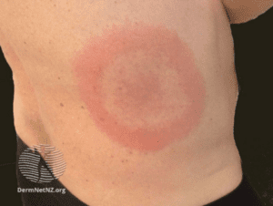
- Erythema migrans (EM)
- 70 to 80% of infected individuals
- At site of tick bite approximately day 7 to 14 days after bite (range 3 to 30 days)
- Expands slowly and develops central clearing
- May have multiple lesions indicating disseminated disease (not multiple tick bites)
- Fever, headache, and fatigue
- Joint and muscle aches
- Swollen lymph nodes

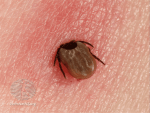
Ixodes tick and Erythema Migrans images courtesy DermNetNZ.org
Early disseminated disease features:
- Neurologic symptoms: lymphocytic meningitis | cranial nerve palsies |radiculopathy | mononeuropathy multiplex | peripheral neuropathy
- Carditis : palpitations, chest pain, light headedness, fainting, shortness of breath, and difficulty breathing with exertion
- Multiple EM lesions
- Conjunctivitis
Late disease features (months to years after tick bite):
- Large joing polyarthralgia
- Confluent mononeuropathy multiplex
- Encephalomyelitis
Testing
Note: The CDC has updated the Lyme laboratory screening algorithm
- CDC still recommends the two step approach
- Step 1: Serologic testing using a sensitive enzyme immunoassay (EIA) or immunofluorescence assay
- Step 2: Follow step one with a western immunoblot assay for specimens yielding positive or equivocal results
- Modified two-step approach
- Two enzyme immunoassays (EIA) are run concurrently or sequentially
- The CDC states that
When cleared by FDA for this purpose, serologic assays that utilize a second EIA in place of western immunoblot assay are acceptable alternatives for the serologic diagnosis of Lyme disease
Note: When EM is present and patient has been to a Lyme endemic area serologic testing is not recommended and treatment can be initiated following clinical diagnosis
KEY POINTS:
- Two-tiered serologic testing is more sensitive the longer infection has been present
- Myth that Lyme disease can become chronic and no longer detectable on lab testing
- Testing can remain positive for years so can not be used to test for cure
Treatment
- First line therapy for adults with early localized Lyme disease
- Doxycycline: 100 mg twice per day for 10 days, or
- Amoxicillin: 500 mg 3 times per day for 14 days, or
- Cefuroxime axetil (500 mg twice per day) for 14 days
- Therapy for early disseminated Lyme disease with neurologic manifestations:
- Doxycycline 100 mg twice per day for 14 to 21 days, or
- Ceftriaxone IV 2 g daily for 14 to 21 days
- Therapy for early disseminated Lyme disease with mild carditis (1st degree AV block with PR interval ≤ 300 milliseconds:
- Doxycyline 100 mg twice per day for 14 to 21 days, or
- Amoxicillin 500 mg three times per day for 14 to 21 days, or
- Cefuroxime 500 mg twice per day orally for 14 to 21 days
- Therapy for early disseminated Lyme disease with severe carditis (symptomatic, 1st degree AV block with PR interval ≥ 300 milliseconds
- If severe carditis (symptomatic, 1st degree AV block with PR interval ≥300 milliseconds, 2nd or 3rd degree AV block) then patient should be admitted for telemetry monitoring and treated with Ceftriaxone IV 2 g daily for 14 to 21 days
- For treatment of late Lyme disease, including arthritis, the antibiotic course is extended to 28 days using the above antibiotics for early localized disease
- The CDC concurs with the following IDSA recommendation that advises strongly against the treatment of chronic Lyme disease (highest level of evidence ‘I’, based on randomized controlled trials) as overtreatment with unnecessary antibiotics may prove fatal (e.g. septic shock, C. difficile, paraspinal abscess and osteomyelitis):
There is no convincing biologic evidence for the existence of symptomatic chronic B. burgdorferi infection among patients after receipt of recommended treatment regimens for Lyme disease. Antibiotic therapy has not proven to be useful and is not recommended for patients with chronic (⩾6 months) subjective symptoms after recommended treatment regimens for Lyme disease.
Prophylaxis: Management of asymptomatic individuals following a tick bite
- Routine use of antimicrobial prophylaxis or serologic testing is not recommended
- A single dose of doxycycline may be offered to adult patients (200 mg dose) when all the following are met:
- The attached tick can be reliably identified as an adult or nymphal I. scapularis deer tick that is estimated to have been attached for ⩾36 h based on the degree of engorgement of the tick with blood or on certainty about the time of exposure to the tick
- Prophylaxis can be started within 72 h of the time that the tick was removed
- Ecologic information indicates that the local rate of infection of these ticks with B. burgdorferi is ⩾20% (occurs in parts of New England, in parts of the mid-Atlantic States, and in parts of Minnesota and Wisconsin)
- Doxycycline is not contraindicated
Tick Removal
- The risk of getting a tick-borne disease is small if the tick is removed soon after it becomes attached
- Deer ticks must remain attached one to two days to transmit Lyme disease, and about one day for other tick-borne diseases
- Use tweezers to grasp the tick close to its mouth
- Gently and slowly pull the tick straight outward
- To avoid contact with the bacteria, if present, do not squeeze the ticks’ body
- Wash the area and apply an antiseptic to the bite
- Watch for early signs and symptoms of Lyme disease
Learn More – Primary Sources:
CDC MMWR: Serious Bacterial Infections Acquired During Treatment of Patients Given a Diagnosis of Chronic Lyme Disease — United States
CDC: Lyme Disease Diagnosis, Treatment and Testing
Clinical Practice Guidelines by the Infectious Diseases Society of America (IDSA), American Academy of Neurology (AAN), and American College of Rheumatology (ACR): 2020 Guidelines for the Prevention, Diagnosis and Treatment of Lyme Disease
Signs and Symptoms of Untreated Lyme Disease
Minnesota Department of Natural Resources: Deer Ticks
CDC: Lyme Disease Transmission
JAMA: Lyme Disease in 2018 – What Is New (and What Is Not)
FDA clears new indications for existing Lyme disease tests that may help streamline diagnoses
CDC: Updated CDC Recommendation for Serologic Diagnosis of Lyme Disease
BMJ State-of-the-Art Review: Lyme borreliosis: diagnosis and management
Prolactinoma: Early Detection, Evaluation and Management
CLINICAL ACTIONS:
Prolactinomas are generally benign prolactin-secreting tumors and account for 40-66% of all pituitary adenomas. The vast majority are microadenomas (diameter < 1cm) and suppress the hypothalamic-pituitary gonadal hormonal axis, while 10% are macroadenomas (≥ 1cm) and may cause additional mass effects due to size. Age prevalence varies widely, but they are commonly found in women during childbearing years, in part due to development of menstrual irregularities. Despite their benign nature, if diagnosis is delayed bone loss and vertebral fractures can occur, and the loss of bone density can be permanent.
Clinical signs and symptoms:
- Oligo or amenorrhea
- Galactorrhea and gynecomastia
- Loss of libido and erectile dysfunction
- Infertility
- Mass effect:
- Headache
- Visual field abnormalities
Evaluation:
- Endocrine Society practice guideline
- History should be obtained to rule out obvious causes of elevated prolactin such as medications
- MRI pituitary with and without contrast to assess size and type of tumor
- Prolactin levels
- Prolactin levels above 200 µg/L is usually a prolactinoma
- Prolactin levels above 500 µg/L likely indicates a macroprolactinoma
- Prolactin macroadenomas can present with a falsely normal prolactin level due to the “hook effect” (false negative levels if excessive amount of analyte is present)
- When prolactin values are lower than expected in a patient, consider discussion with endocrinologist or clinical pathologist for further guidance
- Non-prolactin secreting pituitary adenomas can cause pituitary stalk or hypothalamus compression and consequent prolactinemia
- TSH, free thyroxine (FT4), and creatinine levels to exclude secondary causes
Treatment:
- Minimal symptoms (mild galactorrhea and normal menses): observation and monitor q6-12 months may be acceptable
- Oligo or amenorrhea (pregnancy not desired): oral contraceptives or other estrogen/progesterone combinations
- Most patients placed on dopamine agonists
- Cabergoline > bromocriptine in reducing prolactin levels
- Nearly 80% of patients treated with dopamine agonists will normalize prolactin level and reduce the size of their adenoma
- Transsphenoidal surgery
- Can result in normal prolactin levels in majority of microadenomas and up to 40% in macroadenomas
- Recommended: When dopamine agonists not tolerated/desired | Acute tumor complications |Visual deficits not corrected with medical therapy
- Recurrence is possible (20% over 10 years)
- Radiotherapy
- Rarely used for those cases that do not respond to the above
- Chemotherapy
- Temozolamide rarely used with limited success
- Follow Up
- Once prolactin levels have improved monitoring is recommended with repeat prolactin levels every 3 to 6 months for the first year and then every 6 to 12 months thereafter
- Therapy may be tapered after 2 years of treatment for patients with normal prolactin levels and no visible tumor on MRI
- Recurrence rates after stopping dopamine agonists is between 26 to 69% and most likely to occur in the first year, and should be monitored with serial prolactin measurements every 3 months for the first year and annually thereafter
SYNOPSIS:
The prevalence of prolactinomas is reported to be between 35 to 50 per 100,000. They are most commonly seen in women (10:1 ratio female/male) and the usual age range is between 20 to 50 years of age. Dopamine originating in hypothalamic neurons is a principal inhibitory regulator of prolactin release by pituitary lactotrophs and this pathway is the basis of medical treatments. Fortunately, only a minority of microadenomas will continue to grow (< 10%) but early detection, monitoring and a management plan, which may be multidisciplinary, is required for good outcomes. Consider accessing expertise in endocrinology and radiology to ensure correct differential between prolactinoma and non-secreting pituitary adenoma as treatment for the latter is usually surgical, not medical.
KEY POINTS:
Severe adverse effects of dopamine inhibitors are unusual but cabergoline may include compulsive behavior (e.g. excessive gambling) as well as cardiac valvular abnormalities at high doses
- Other causes of hyperprolactinemia include
- Pituitary disorders (e.g. Cushing disease)
- Hypothalamic disorders (e.g. non-secreting pituitary adenoma)
- Neurogenic (e.g. chest wall or spinal cord lesions)
- Medical
- Hypothyroidism
- Chronic kidney disease
- Cirrhosis
- Medications
- Antipsychotics (e.g. phenothiazines)
- Antidepressants (e.g. tricyclics, MAOIs, SSRIs)
- Antihypertensives (e.g. verapamil, labetolol)
- Anticonvulsants (e.g. phenytoin)
- Prokinetics (e.g. metoclopramide)
- Hormones (e.g. estrogen)
- H2 blockers (e.g. cimetidine, ranitidine)
- Controlled substances (e.g. opiates, cocaine, marijuana)
- Other (e.g. alcohol)
Learn More – Primary Sources:
Orphanet J Rare Disease: The risks of overlooking the diagnosis of secreting pituitary adenomas
Diagnosis and Treatment of Pituitary Adenomas: A Review
Guidelines of the Pituitary Society for the diagnosis and management of prolactinomas
Diagnosis and Treatment of Hyperprolactinemia: An Endocrine Society Clinical Practice Guideline
Prolactinoma Management
The epidemiology, diagnosis and treatment of Prolactinomas: The old and the new
Underappreciation of the “Hook Effect” leading to Mismanagement of a Patient, a Tale of Preventable Disaster
Screening & Treatment of Gynecologic infections in the HIV-Positive Woman
Gynecologic infections are more common, and may be more difficult to eradicate, in the HIV population. Overall, treatment protocols remain the same, irrespective of HIV status, although there are some differences depending on the disorder.
CLINICAL ACTIONS:
- Screen at entry to care and at least annually for the following: N. gonorrhoeae, C. trachomatis, syphilis and vaginal trichomoniasis
- Screen for hepatitis C on entry to care
- At-risk seronegative individuals should be screened at least annually
- Consider type specific HSV serologic testing for those presenting for an STD evaluation
- Approximately 70% of persons with HIV are HSV-2 seropositive | 95% are seropositive for either HSV-1 or HSV-2
- HSV-2 infection increases the risk of HIV acquisition two- to three-fold and in coinfected patients
- HSV-2 reactivation results in increases in HIV RNA levels in blood and genital secretions
- Screen on entry to care for hepatitis B with HBsAg, anti-HBc and/or anti-HBs
- Offer vaccination to seronegative individuals with hepatitis B or combined hepatitis A and B vaccine
- Recheck immunity after vaccination complete
SYNOPSIS:
While it is critical to remain vigilant with regard to STDs and pelvic infections, women with ulcerative conditions of the genitalia, including syphilis and herpes, are at increased risk of HIV acquisition and transmission to partners, lending an urgency to prompt treatment or suppression (see separate entry on ulcerative conditions).
KEY POINTS:
- Bacterial vaginosis is more prevalent/persistent in HIV-positive women
- Diagnosis and treatment options are the same
- Vulvovaginal candidiasis is more common among HIV-positive women and associated with decreased CD4+ counts
- Treatment is the same as for HIV-negative women
- For azole-refractory Candida glabrata vaginitis
- Boric acid 600 mg vaginal suppository once daily for 14 days
- Note: Severe or recurrent vaginitis should be treated with oral fluconazole (100 to 200 mg) or topical antifungals for ≥7 days
- Treatment for gonorrhea/chlamydia is the same as for HIV-negative women
- Retest 3 months after treatment as reinfection is common
- Pelvic inflammatory disease is treated with the same antimicrobials for the same duration as for HIV-negative
- There is a greater incidence of tubo-ovarian abscess among HIV-positive, but overall response to therapy is the same as for HIV-negative
- Trichomoniasis is more prevalent among HIV-positive and should be treated with a one week course of metronidazole
- Parasitic conditions such as scabies or pediculosis pubis are treated the same regardless of HIV status
Learn More – Primary Sources:
ACOG Practice Bulletin 167: Gynecologic Care for Women and Adolescents with Human Immunodeficiency Virus
NIH Guidelines for Prevention and Treatment of Opportunistic Infections in HIV-Infected Adults and Adolescents
CDC: Sexually Transmitted Infections Treatment Guidelines 2021
Ulcerative Genital Conditions in the HIV-Positive Woman
Genital, anal, or perianal ulcers are generally caused by syphilis or herpes. Chancroid, lymphogranuloma venereum (LGV), and granuloma inguinale (donovaniasis) are less likely diagnoses; however, all these conditions are associated with increased HIV acquisition and transmission. Guidelines for screening and treatment differ in some cases from those for the HIV-negative patient.
CLINICAL ACTIONS:
- Evaluate first for herpes and syphilis first
- Other causes such as chancroid and lymphogranuloma venereum should then be considered
Herpes Simplex (HSV)
Syphilis
Chancroid
Lymphogranuloma Venereum (LGV)
Granuloma Inguinale (donovaniasis)
Herpes Simplex lesions:
- Caused by herpes simplex viruses (HSV)
- Common and may be severe, painful and atypical
- Offer HSV type-specific serologic testing on initial HIV evaluation for asymptomatic individuals
- HSV DNA polymerase chain reaction (PCR) and viral culture are preferred methods for diagnosis of mucocutaneous HSV lesions caused by HSV
- Consider suppression or episodic therapy to decrease clinical manifestations if HSV-2 is detected
- Does not reduce risk for HIV transmission or HSV-2 transmission to susceptible sex partners
- Generally higher doses/longer duration of treatment are recommended for HIV-positive women
Daily suppression
- Acyclovir 400-800 mg BID or TID
or
or
Episodic flares
- Acyclovir 400 mg TID x 5-10 days
or
- Valacyclovir 1 g BID x 5-10 days
or
- Famcycolovir 500 mg BID x 5-10 days
Syphilis:
- Systemic disease caused by Tremonema Pallidum
- Primary syphilis is characterized by genital ulcers, though secondary/ tertiary/latent forms are not
- A presumptive diagnosis of syphilis requires a treponemal test (i.e fluorescent treponemal antibody absorbed tests [FTA-ABS]) plus a nontreponemal test (i.e. Venereal Disease Research Laboratory [VDRL] or Rapid Plasma Reagin [RPR])
- Screen for syphilis on initial HIV evaluation and annually thereafter
- Treatment is the same regardless of HIV status
- Penicillin G IM is the preferred treatment for all stages; the preparation (benzathine, aqueous procaine aqueous crystalline), dosage and length of treatment depend on the stage
Chancroid:
- Painful, genital ulcers with suppurative lymphadenopathy caused by ducreyi
- Treatment is the same regardless of HIV status
- Close follow up is required as treatment failure is increased in the HIV positive
Treatment
or
or
- Ciprofloxacin 500 mg po BID x 3 days
or
- Erythromycin 500 mg tid x 7 days
Lymphogranuloma Venereum (LGV)
- Tender, unilateral groin/femoral lymphadenopathy with a self-limited genital ulcer/papule caused by C. trachomatis serovars L1, L2, or L3
- Proctitis may occur
- Diagnose by identifying C. trachomatis from the lesion by culture, direct immunofluorescence or nucleic acid detection
- Treatment is the same regardless of HIV status
Treatment
- Doxycycline 100 mg BID x 3 weeks
or
- Erythromycin base 500 mg QID x 3 weeks
Granuloma Inguinale (Donovanosis):
- Painless ulcerative disease characterized by beefy red, highly vascular lesions caused by Klebsiella granulomatis
- Diagnose by identifying dark-staining “Donovan Bodies” on biopsy or tissue
- Treatment is the same regardless of HIV status
Treatment
- Azithromycin 1 gm once/week or 500 mg/day x 3 weeks/until lesions healed
or
- Doxycycline 100 mg BID X 3 weeks/until lesions healed
or
- Ciprofloxacin 750 mg BID x 3 weeks/until lesions healed
or
- Erythromycin base 500 mg QID x 3 weeks/until lesions healed
or
- Trimethoprim-sulfamethoxazole double strength BID x 3 weeks/until healed
SYNOPSIS:
Vaginal, vulvar, and sexually transmitted infections may increase the risk of HIV transmission. Prompt diagnosis and treatment decreases the likelihood of transmission; public health standards require providers to presumptively treat any patient, regardless of HIV status, with suspected syphilis immediately and before test results are available. Presumptive treatment of primary herpes is also recommended as early treatment maximizes success.
KEY POINTS:
- Genital, anal, or perianal lesions may not necessarily be infectious (e.g. trauma, carcinoma, aphthous, drug eruption, psoriasis)
- Medical history and physical exam findings are frequently inaccurate and should be followed by tests that reflect conditions prevalent in the area
- Consider biopsy for ulcers not responding to therapy or if the diagnosis is unclear
- If HIV status is unknown, HIV testing should be offered to any woman with genital/anal ulcers presenting for treatment.
Diagnosis codes:
- D28.9 benign neoplasm of female genital organs, unspecified
- Z21 HIV infection
Learn More – Primary Sources:
2015 Sexually Transmitted Disease Treatment Guidelines
ACOG Practice Bulletin 167: Gynecologic Care for Women and Adolescents with Human Immunodeficiency Virus
NIH Guidelines for Prevention and Treatment of Opportunistic Infections in HIV-Infected Adults and Adolescents