Microarrays and Microdeletions: Key Concepts Summarized
WHAT IS IT?
A microarray describes a newer technology that can identify small duplications or deletions of genetic material that previously could not be identified using conventional karyotyping alone. It has become a critical tool to help identify submicroscopic chromosomal deletions/duplications that underlie clinically significant syndromes in the prenatal period and throughout the lifespan.
Key Concepts
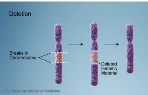
What is a Deletion?
A deletion describes a chromosomal break where genetic material is lost
Credit: US National Library of Medicine
Deletions can be large or small, and can occur anywhere along a chromosome
Terminal deletion (end of a chromosome)
Interstitial (within the chromosome)
Duplication describes when additional material is gained
What is a Microdeletion?
Definition is based on size of missing DNA sequence
1Mb (megabase) = 1 million base pairs
Conventional karyotype is based on light microscopy and can usually only detect deletions > 5 Mb
Microdeletions refer to deletions smaller than those that can be seen on karyotype – i.e., < 5 Mb
What is a CNV (copy number variant)?
Deletions or duplications are ≥ 1 kb (1000 base pairs) to many hundreds of Kb in size
Small CNVs are common and found in 5 to 10% of individuals in the general population
Only 1 to 2% will have CNVs > 1Mb
Most CNVs are simply a part of normal human variation
If CNVs delete or add sufficiently large number of genes or result in a breakpoint within a gene, they may have significant clinical consequences
Key Points:
CGH vs SNP arrays
CGH (Comparative genome hybridization)
The array is constructed with single short DNA sequences from the regions of interest
‘Test’ and ‘control’ DNA are labelled with different fluorescent colors and then hybridized (annealed) to complementary sequences of interest on the array
The relative amounts of test vs control DNA can be measured
Excess control DNA signal color signifies a deletion in the test sample
Excess test DNA signal color signifies duplication in the test sample
SNP array
The array is constructed with short DNA sequences
Each region of interest will have two DNA sequences, representing the two possible alleles (versions) of known SNPs
Only ‘test DNA’ is labelled and hybridized to the allele specific probes within the array
Relative intensity of the hybridization signal is used to detect if DNA sequence is deleted or duplicated
Low/absent intensity signal signifies deletion
Excess signal signifies duplication
Additional capabilities of SNP array compared to CGH
Can detect triploidy, maternal cell contamination and mosacism
Can detect consanguinity and uniparental disomy (UPD) which can be associated with genetic syndromes
Rather than having one copy of each SNP allele, a region of identical SNPs, known as absence of heterozygosity (AOH), will be apparent
Labs may report AOH, raising concern for
Autosomal recessive disorders related to genes in that particular sequence or
The Use of Microarrays in the Prenatal and Postnatal Setting – Current Professional Guidelines
CLINICAL ACTIONS:
Chromosomal microarray (also known as CMA) technology has revolutionized the practice of genetics and is now commonly ordered prenatally and throughout the lifespan. Structural chromosomal anomalies, once too small to be ‘seen’ using conventional karyotyping, can now be detected using molecular techniques. Microarrays can detect copy number variants (CNVs) and therefore identify chromosomal regions where there is excess DNA (a duplication) or missing DNA (a deletion).
Consider the following when counseling:
Benefits of Microarrays
Prenatal detection of clinically significant CNVs that would have been missed using conventional karyotyping alone
6% of abnormal fetuses with a normal karyotype may have pathogenic CNVs or likely pathogenic CNVs
Isolated finding: 5.6%
Multiple anomalies: 9.1%
1.7% of normal fetuses and normal karyotype may have pathogenic CNVs or likely pathogenic CNVs
Postnatal detection of significant CNVs that would have been missed using conventional karyotyping alone
Developmental delay and intellectual disability
An additional 12.2% – 19% pathogenic anomalies May be detected with the addition of microarray
Can be performed on tissue that is no longer viable
If DNA is present and of sufficient quality, test can be run on stillbirth specimens or products of conception
Limitations of Microarrays
Balanced translocations
Balanced translocations occur in approximately 1/500 individuals and are usually benign
Because there is minimal/no additional or deleted genetic material, balanced translocations will go undetected with microarray but depending on size, may be seen with conventional karyotyping
Serious consequences are still possible with balanced translocations due to breakpoints disrupting genes
ACMG (2018) notes the importance of balanced rearrangements (e.g., translocation, inversion, insertion) and states that “recurrence risk counseling is indicated for offspring of parent with a balanced rearrangement
Estimated additional diagnostic yield of 0.78 to 1.3% if a G-banded karyotype is performed following a negative microarray
Single gene disorders
May be caused by a single or small base pair change and will not be detected on microarray
Variants of uncertain significance (VUS)
Can occur in 1 to 2% of cases
May cause parental anxiety and will require additional expert counseling and follow-up
Over time, VUSs are being categorized as benign or pathogenic as additional reports are incorporated into databases
SYNOPSIS:
The data from a major NICHD study in 2012 provided the support for introducing microarray technology into prenatal clinical care, with previous studies highlighting the benefits in the postnatal period and beyond. Similar to a conventional karyotype, microarrays can detect aneuplodies and larger structural chromosomal changes. In addition, microarray technology allows for the identification of small duplications and microdeletions that would otherwise go unreported. There are limitations and expert counseling is required to provide optimal care and informed decision making.
KEY POINTS:
Prenatal Recommendations
SMFM
Fetal structural anomalies on prenatal ultrasound or stillbirth
Microarray replaces conventional karyotype (1A)
Patient undergoing invasive testing and no anomalies identified
Both options, conventional karyotype and microarray, should be discussed (1B)
SOGC & CCMG
Microarray should be offered following a normal rapid aneuploidy screen when
Multiple fetal malformations are detected (II-1A) or
NT ≥3.5 mm (II-2B)
Postnatal Recommendations
ACMG
Microarray is a first-line test in the initial postnatal evaluation in the following clinical scenarios
Multiple anomalies not specific to a well-delineated genetic syndrome
Should Amniocentesis or Chorionic Villus Sampling Be Offered to All Pregnant Women?
CLINICAL ACTIONS:
Invasive prenatal diagnostic testing usually refers to amniocentesis (analysis of amniotic fluid cells) or chorionic villus sampling (placental cells). Prenatal healthcare providers should
Offer all patients the option of prenatal invasive testing or prenatal screening
Counsel patients that unlike invasive tests which cover all chromosomal abnormalities, traditional screening tests or even the newer cfDNA (NIPS / NIPT) tests will only screen for specific and limited chromosomal abnormalities
although cfDNA has superior detection for Down syndrome, traditional first-trimester combined screening can detect additional structural and anatomical anomalies because of the ultrasound component of the test
Identify the following risk factors for fetal aneuploidy and consider referral to genetic counseling and high risk OB services if patient requires more in depth counseling or has additional concerns
Maternal age of 35 or older at EDD: the risk for chromosomal aneuploidy increases throughout the reproductive lifespan, not just after age 35
If either parent of the fetus has an unusual chromosome makeup or aneuploidy, such as an additional X or Y chromosome
ACOG states
A patient’s baseline risk for chromosomal abnormalities should not limit testing options; serum screening with or without NT ultrasound or cell-free DNA screening and diagnostic testing (CVS or amniocentesis) should be discussed and offered to all patients regardless of maternal age or risk for chromosomal abnormality
ACMG recommends
Allowing patients to select diagnostic or screening approaches for the detection of fetal aneuploidy and/or genomic changes that are consistent with their personal goals and preferences
Informing all pregnant women that diagnostic testing (CVS or amniocentesis) is an option for the detection of chromosome abnormalities and clinically significant CNVs
SYNOPSIS:
The cells obtained from amniocentesis or CVS are analyzed to determine if the number of chromosomes are correct (46) and whether there are structural changes such as deletions or duplications. Routine karyotyping is done using light microscopy. If changes are smaller than the resolution of a microscope, then molecular techniques are required and these small alterations are called microduplications or microdeletions (see ‘Related ObG Topics’ below). Presently, despite major advances in screening technologies, diagnosis of fetal aneuploidy still requires an invasive test.
KEY POINTS:
Miscarriage Risks
In expert hands, there is a 0.1 to 0.3% chance of miscarriage associated with invasive prenatal testing
Cochrane Review (Alfirevic et al., 2017) has released its review on amnio/CVS safety
2nd trimester amnio increased risk of pregnancy loss, but it was not possible to quantify the loss rate, based on one study that is now over 30 years old
Early amnio (11w0d-12w6d) is associated with pregnancy loss and clubfoot compared to 2nd trimester amnio (15w0d-16w6d)
Transcervical CVS may be associated with higher loss rate compared to 2nd trimester amnio but the quality of the evidence was downgraded due to heterogeneity between studies
Wulff et al. (Ultrasound Obstet Gynecol, 2016)
Using propensity scoring on a nationwide database of approximately 150,000 women, did not find an increased risk of miscarriage or stillbirth due to amnio or CVS when indications for the procedures were taken in to account (see Related OBG Topics below for review of this paper and other recent papers on this subject of procedure related fetal loss)
Salomon et al. (Ultrasound Obstet Gynecol, 2019)
Estimated procedure-related risk of miscarriage after amniocentesis and chorionic villus sampling (CVS)
Performed a systematic review and meta-analysis, covering 20 controlled studies
The authors concluded
…amniocentesis is associated with a procedure-related risk of 1:300 at most, or more likely, no significant increase in risk
With regard to CVS, our results demonstrate that, there is no significant procedure-related risk associated with undertaking this procedure
Routine Karyotyping or Microarray?
Abnormal prenatal ultrasound with structural abnormality
A chromosomal microarray analysis that can detect submicroscopic changes is recommended
Standard karyotype may miss 6% of important chromosome changes
Normal prenatal ultrasound
A chromosomal microarray can be offered because 1.7% of significant chromosome changes will not be detected on a standard karyotype approach
Microarray limitations to discuss with patients (see ‘Related ObG Topics’ below for more on the benefits and limitations of microarray analysis)
In a small number of cases, the laboratory may identify copy number variants of uncertain significance (VUS), also referred to as variants of uncertain significance (VOUS)
Over time, as databases grow, VUSs can be re-categorized as benign or pathogenic
Microarrays cannot detect low levels of mosaicism (more than one cell line) or balanced translocations
Small risk that while overall DNA appears balanced on a microarray, the breaks involved in the translocation may have disrupted a gene and lead to abnormal protein production
Additional Considerations
A patient who would not terminate a pregnancy
Important information that may impact the management of a pregnancy may be obtained on invasive testing beyond termination of pregnancy
Therefore, all patients should be offered the option of screening or invasive testing and have the option of accepting or declining testing irrespective of future reproductive choices
Diagnosis code: diagnosis code will vary depending on indication
Procedure codes: amniocentesis- 59000; sono guidance for amniocentesis- 76946
Procedure codes: CVS- 59015; sono guidance for CVS-76945
OBG Project CME requires a modern web browser (Internet Explorer 10+, Mozilla Firefox, Apple Safari, Google Chrome, Microsoft Edge). Certain educational activities may require additional software to view multimedia, presentation, or printable versions of their content. These activities will be marked as such and will provide links to the required software. That software may be: Adobe Flash, Apple QuickTime, Adobe Acrobat, Microsoft PowerPoint, Windows Media Player, or Real Networks Real One Player.
Disclosure of Unlabeled Use
This educational activity may contain discussion of published and/or investigational uses of agents that are not indicated by the FDA. The planners of this activity do not recommend the use of any agent outside of the labeled indications.
The opinions expressed in the educational activity are those of the faculty and do not necessarily represent the views of the planners. Please refer to the official prescribing information for each product for discussion of approved indications, contraindications, and warnings.
Disclaimer
Participants have an implied responsibility to use the newly acquired information to enhance patient outcomes and their own professional development. The information
presented in this activity is not meant to serve as a guideline for patient management. Any procedures, medications, or other courses of diagnosis or treatment discussed or suggested in this activity should not be used by clinicians without evaluation of their patient’s conditions and possible contraindications and/or dangers in use, review of any applicable manufacturer’s product information, and comparison with recommendations of other authorities.
Jointly provided by
NOT ENOUGH CME HOURS
It appears you don't have enough CME Hours to take this Post-Test. We no longer offer Hours.
Leaving ObG Website
You are now leaving the ObG website and on your way to PRIORITY at UCSF, an independent website. Therefore, we are not responsible for the content or availability of this site