For Physicians. By Physicians.
For Physicians. By Physicians.

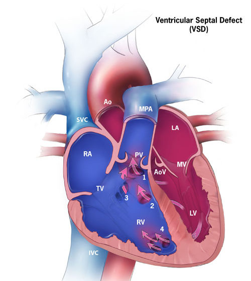
Image credit: Centers for Disease Control and Prevention, National Center on Birth Defects and Developmental Disabilities
AHA Circulation Journal Review: Ventricular Septal Defects
CDC: Facts About Ventricular Septal Defect

Tetralogy of Fallot (TOF) is a congenital heart defect, that occurs in approximately 1 in every 2,500 live births and accounts for 7-10% of all congenital heart defects
TOF is comprised of 4 major abnormalities:
Cause is usually unknown, but may be associated with genetic issues
Categorized as a ‘critical congenital heart defect’, which means a likelihood of surgery and intensive management and procedures during the first year of life

Credit: Mariana Ruiz Villarreal
CDC: Facts about Tetralogy of Fallot
CDC: Facts about Critical Congenital Heart Defects
CDC: Improved National Prevalence Estimates for 18 Selected Major Birth Defects
ISUOG Practice Guidelines (updated): sonographic screening examination of the fetal heart
ACOG Practice Bulletin No. 162: Prenatal Diagnostic Testing for Genetic Disorders

In a normal heart, the pulmonary artery carries deoxygenated blood to the lungs. Oxygenated blood returns to the left side of the heart and the aorta then pumps the oxygenated blood to the rest of the body. In Transposition of the Great Arteries (TGA), the pulmonary artery and aorta have changed places (i.e., they are transposed). Therefore:
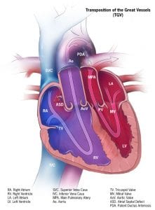
Image credit: Centers for Disease Control and Prevention, National Center on Birth Defects and Developmental Disabilities
CDC: Facts about dextro-Transposition of the Great Arteries (d-TGA)
Circulation-AHA journal: Transposition of the Great Arteries
CDC: Facts about Critical Congenital Heart Defects
Current diagnosis and treatments for critical congenital heart defects
Fetal Growth and Neurodevelopmental Outcome in Congenital Heart Disease
Maternal Fetal Medicine Specialist Locator-SMFM

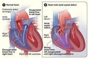
Image credit – National Heart Lung and Blood Institute (NIH)
ASD is typically sporadic, however may occur as an isolated finding in families or as part of various genetic syndromes including but not limited to Down syndrome, DiGeorge syndrome and Holt-Oram syndrome which involves variable radial ray anomalies.
AHA Circulation Journal -Atrial Septal Defects in the Adult
CDC: Facts about Atrial Septal Defect
ACOG Practice Bulletin No. 162: Prenatal Diagnostic Testing for Genetic Disorders
Please log in to ObGFirst to access this page
OBG Project CME requires a modern web browser (Internet Explorer 10+, Mozilla Firefox, Apple Safari, Google Chrome, Microsoft Edge). Certain educational activities may require additional software to view multimedia, presentation, or printable versions of their content. These activities will be marked as such and will provide links to the required software. That software may be: Adobe Flash, Apple QuickTime, Adobe Acrobat, Microsoft PowerPoint, Windows Media Player, or Real Networks Real One Player.
This educational activity may contain discussion of published and/or investigational uses of agents that are not indicated by the FDA. The planners of this activity do not recommend the use of any agent outside of the labeled indications.
The opinions expressed in the educational activity are those of the faculty and do not necessarily represent the views of the planners. Please refer to the official prescribing information for each product for discussion of approved indications, contraindications, and warnings.
Participants have an implied responsibility to use the newly acquired information to enhance patient outcomes and their own professional development. The information
presented in this activity is not meant to serve as a guideline for patient management. Any procedures, medications, or other courses of diagnosis or treatment discussed or suggested in this activity should not be used by clinicians without evaluation of their patient’s conditions and possible contraindications and/or dangers in use, review of any applicable manufacturer’s product information, and comparison with recommendations of other authorities.
Jointly provided by