For Physicians. By Physicians.™

ObGFirst: Get guideline notifications, fast. First month free!Click here

Basal Cell Carcinoma: Clinical Presentation and Management

SUMMARY:

Basal cell carcinoma (BCC) is the most common form of invasive skin cancer in the world and affects more than 3.3 million persons annually in the United States. 85% of BCCs occur on the head and neck region, due to UV light exposure and can be readily treated as outpatient if detected early. While they rarely become metastatic, BCC can be locally destructive and cause significant disfigurement. Recently, an expert work group was convened to determine evidence-based guidelines to provide guidance for biopsy, staging, treatment and follow-up of BCC.

Background

BCC is an extremely common skin cancer | Main risk factor for development of BCC is ultraviolet light exposure | Many BCCs occur in relatively sun-protected sites, such as behind the ears

  • Arises from basal keratinocytes of the epidermis
  • Light skinned individuals are at higher risk | BCCs in skin of color are rare
  • When diagnosed early, the majority of BCCs can be readily treated in the office
  • Severe disfigurement can occur when not found and treated early
  • Recurrence is common if initially inadequately treated

Clinical Presentation

There are 5 clinical types or presentations of BCC: Nodular | Pigmented | Sclerosing/Morpheaform | Superficial | Nevoid

Nodular BCC

  • Most common, classically presents as a pearly white or pink dome-shaped papule with telangiectatic vessels | A bleeding or scabbing sore that heals and recurs | Often becomes ulcerated Cystic | BCC is a variant of nodular BCC and appears as a smooth, round, cystic mass

Pigmented BCC

  • Pigmented BCC contains melanin that will impart a brown, black or blue color through all or part of the lesion | Clinically may resemble a melanoma with a raised border

Sclerosing or Morpheaform BCC

  • Has innocuous superficial characteristics that can mask the potential for deep and wide extension | Appears as a waxy firm flat lesion resembling a scar | Nodules can become apparent later in the course | Margins are generally poorly defined

Superficial BCC

  • Shallow pink or flesh colored plaques often present on the trunk | More common in men | Typically friable and bleed with minor trauma | Expand slowly 

Nevoid BCC Syndrome (Gorlin-Goltz Syndrome)

  • Rare disorder inherited as an autosomal dominant trait | Multiple BCC lesions appearing at birth or in early childhood | Jaw odontogenic keratocysts | Palmar and plantar pits | Lamellar calcification of the falx cerebri | Cleft lip/palate

BCC Risk Factors

  • Exposure to UVB radiation
  • Fair skin, blond hair and light-colored eyes (skin type I)
  • Transplant patients have 10-100 times greater risk than the general population Previous BCC occurrence
  • Chronic arsenic exposure 
  • Ionizing radiation therapy  

Differential Diagnosis

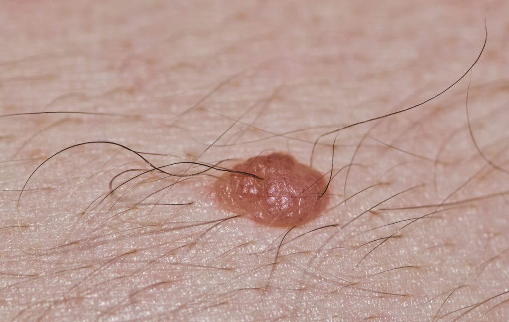
Dermal Nevus


Sebaceous Hyperplasia


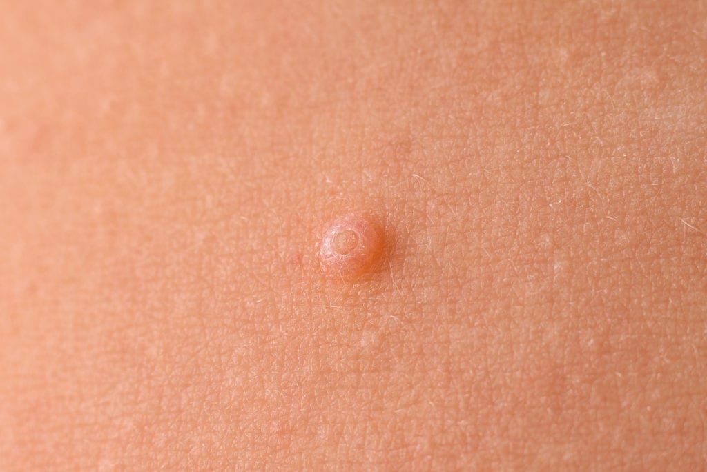
Molluscum Contagiosum

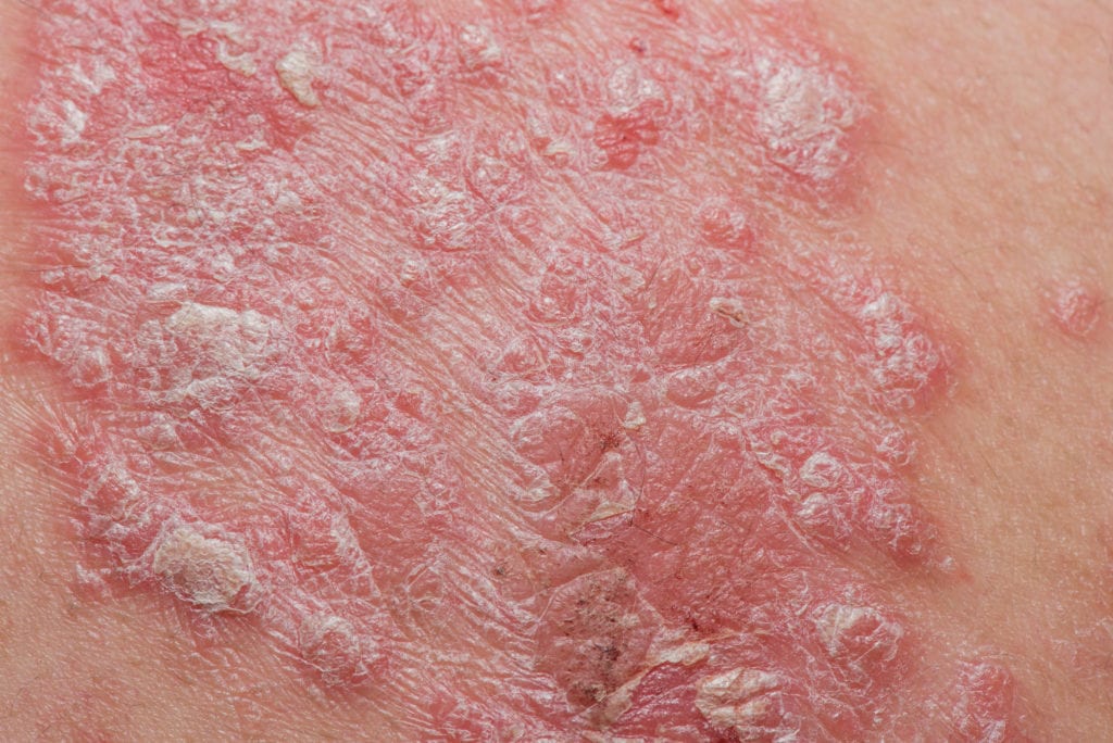
Psoriasis

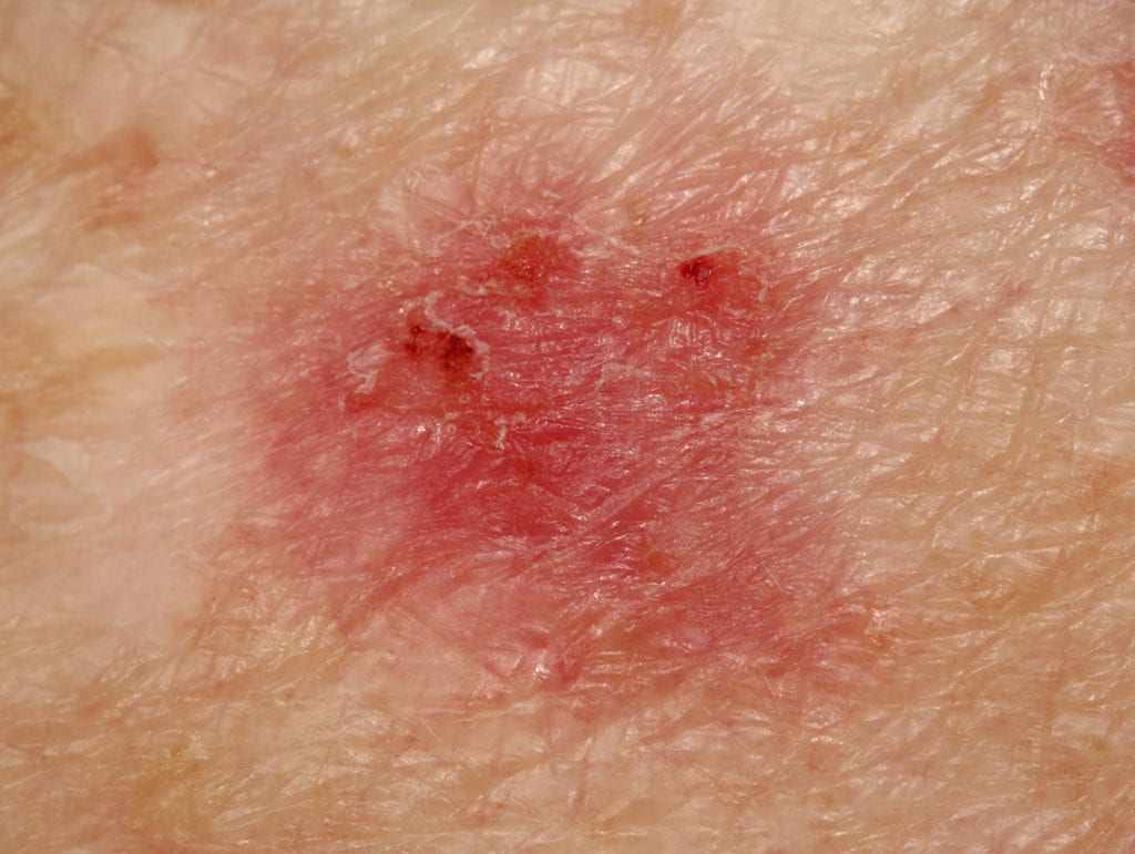
Extramammary Paget’s or Bowen’s Disease
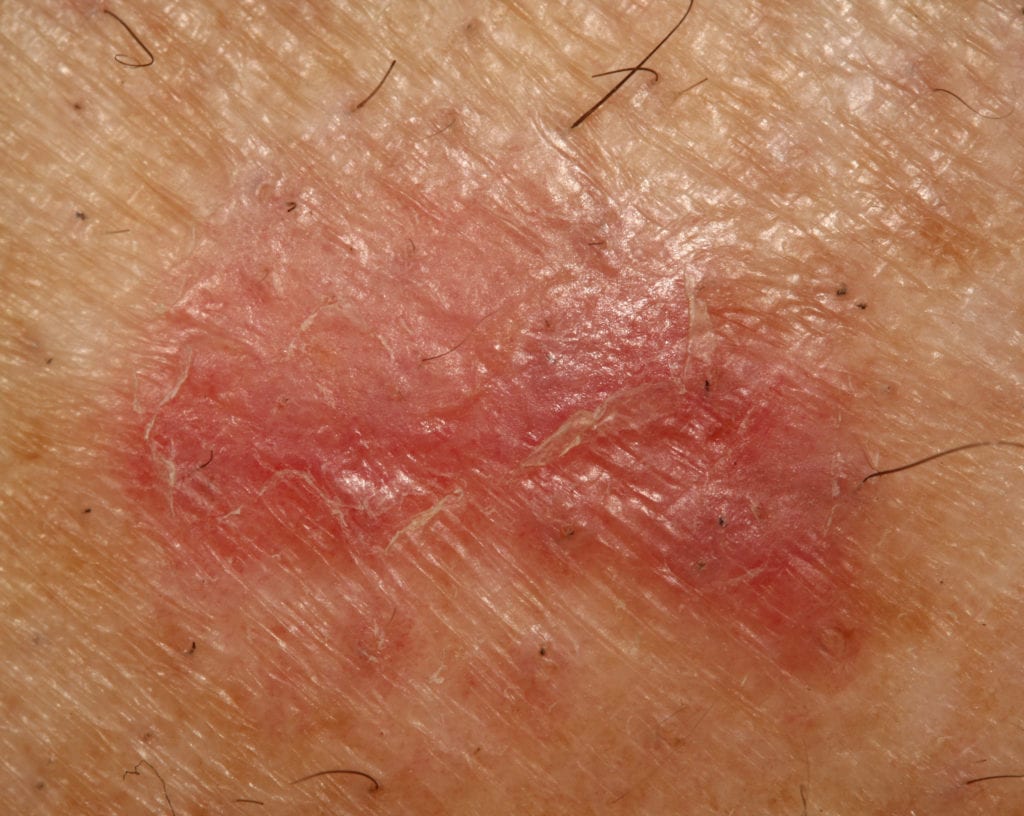

Squamous Cell Carcinoma
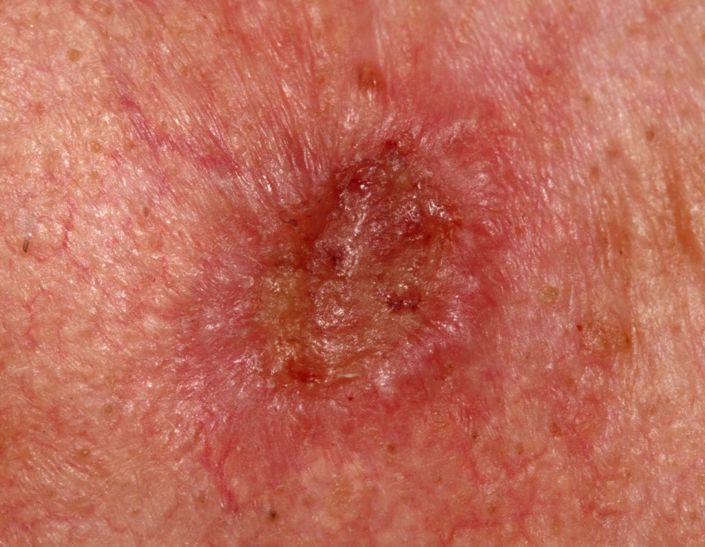

Melanoma (can look similar to pigmented BCC)


Evaluation and Management 

A formal staging system for risk stratification for patients with BCC is not available

  • When metastasis occur the same TNM classification used for SCC, established by the American Joint Committee of Cancer, is also applied to BCC 
  • The most useful stratification of BCC is provided by the National Comprehensive Cancer Network Guidelines
    • Low- versus high-risk is based on: Location | Size | Borders | Primary or recurrent | Immunosuppression | Site of prior radiation therapy | Pathology

The available literature does not identify a single optimal biopsy technique, with either saucerization punch or excisional biopsy with margins acceptable

Treatment Options 

Treatment options include both surgical and non-surgical, with surgery remaining the first-line treatment choice

Surgical Treatment

  • Electrodessication and curettage for low-risk tumors
  • Standard excision with a 4 mm margin of uninvolved skin and depth to the mid-subcutaneous adipose tissue for low-risk tumors
  • MOHS micrographic surgery for primary and recurrent high-risk BCC not located on the trunk or extremities 

Non-surgical Treatment

  • Cryosurgery may be considered for low-risk BCC when more effective therapies are contraindicated or impractical
  • Topical therapies including imiquimod or 5-FU
  • Photodynamic Therapy (PDT) with 5-ALA (aminolevulinic acid) or MAL (methylaminolevulinate) Laser therapy
  • Radiation therapy in the elderly if they cannot tolerate minor surgical procedures

Metastatic BCC

  • Metastatic BCC is uncommon
    • Occurs via lymphatic spread to regional nodes and hematogenous spread to lung and bone
    • There is no curative chemotherapy for metastatic BCC 
  • Few options are available
    • Vismodegib (hedgehog pathway inhibitor) – first systemic therapy approved by the FDA
    • Platinum based chemotherapy may be considered
    • Radiation to regional node areas may be considered
    • Palliative Care consult

Follow-up and Reducing Risk for Future Skin Cancers

  • Once BCC has been diagnosed, in-office screening for new primary skin cancers (BCC, SCC and melanoma) should be performed at a minimum annually
  • Patient and family should be educated to perform skin self-examinations
  • Sun protection and prevention is key
    • Broad-spectrum sunblock and broad brimmed head coverings to protect skin from UV damage
  • Avoid topical or oral retinoids
  • Avoid dietary supplementation with selenium or beta-carotene
  • Recurrence of BCC can occur many years out from the primary lesion with 18% of recurrences occurring after 5 years 

Primary Sources – Learn More:

American Academy of Dermatology: Guidelines of care for the management of basal cell carcinoma

DermNet NZ

NIH NCI: Skin Cancer Treatment (PDQ®) Health Professional Version

Clinical variants, stages, and management of basal cell carcinoma