Rate of uterine rupture in TOLAC (ACOG PB 205 and additional ‘Primary Sources’ section below)
After 1 LTCS: 0.5 to 0.9%
≥2 LTCS: 0.9% to 3.7%
Previous high vertical cesarean delivery: 10%
Unknown scar (judge based on likelihood of LTCS): 2 to 5%
These risk estimates do not change with successive TOLAC or history of successful VBAC
The safest mode of delivery after cesarean is a successful VBAC, the second safest is elective repeat cesarean (ERCD) and highest risk, both maternal and neonatal, is associated with a cesarean after failed TOLAC (ACOG PB 205)
Appropriate safety mechanisms should be in place (in-house anesthesia and attending physician capable of an emergent C/S) before offering TOLAC routinely
Women with a history of 1 to 2 previous LTCS are appropriate candidates for TOLAC. While risk of rupture is slightly higher in women with 2 previous C/S the rates of successful VBAC are similar in large studies
Based on current recommendation cervical ripening in TOLAC should not include the use of prostaglandins
Perinatal Risks (AHRQ Evidence Reports, 2010)
Risk of maternal death
TOLAC: 0.0019% | ERCD: 0.0096%
Risk of neonatal death
TOLAC: 0.11% | ERCD: 0.06%
An interdelivery interval of <18 months is associated with increased risk of uterine rupture, but is not considered an absolute contraindication (Bujold and Gauthier. Obstet Gynecol, 2010)
BACKGROUND:
Between 1970 and 2016 the rate of cesarean delivery in the US rose from 5% to 31.9% (ACOG PB 205)
This is attributed primarily to the introduction of electronic fetal monitoring and the reduction in both operative vaginal delivery and breech vaginal delivery
The purpose of decreasing both primary and repeat cesarean rates is to combat the increased risks associated with subsequent, multiple cesarean deliveries, most notably
Short term risks: Hemorrhage | Thromboembolism | Infection | Bowel or bladder injuries | Hysterectomy | Abnormal placentation and its associated risks (e.g. PPH, PTD, DIC etc.)
Distinguishing the level and direction of the previous cesarean scar is important due to the fundamental differences in uterine muscle between the lower uterine segment (LUS) (sometimes referred to as the isthmus in research literature) and the upper portion
In the LUS, the myometrium is composed of less contractile horizontally arranged fibers that dilate and stretch during the labor process
The myometrium in the upper uterus is composed of highly contractile fibers that run horizontally, longitudinally and in spiral or mixed directions
These differences lead to vastly different risks when the muscles are exposed to the forces of labor after healing and forming scar
Previous 2- vs 1-layer closure and TOLAC
Current recommendation is for a 2-layer closure for women who may want to TOLAC in future pregnancy
Data does not definitively support 2-layer versus 1-layer, or whether locked versus unlocked repair of the first layer definitively impact rupture risk; therefore, these factors should not be used to determine who is an appropriate candidate
IOL is acceptable in TOLAC, however it does slightly increase the risk of rupture
In a large multicenter study of over 33,000 women the rates for rupture with IOL were (Landon et al. NEJM, 2004)
Induction with any prostaglandin (+/- oxytocin): 1.4%
Induction with oxytocin alone: 1.1%
Spontaneous labor: 0.4%
It is unclear from this and other studies whether it is the length of the induction or whether starting from an unfavorable versus favorable cervix that influences the risk of rupture
There is indication of a dose response between oxytocin and risk of rupture, but there is no clear maximum safe level that has been determined
MFMU VBAC Predictive Calculator
Several VBAC success calculators have been proposed based on large database studies
Vyas et al. (NEJM, 2020) note that race-adjusted VBAC calculators may exacerbate disparities, leading to further increased cesarean birth rates in the nonwhite US population
The MFMU Network VBAC Calculator (see references below) has been updated and no longer includes race or ethnicity
Data derived from Grobman et al. (AJOG, 2021)
While these calculators can be used for shared decision making, there is currently no evidence that VBAC predictive models/tools improve outcomes
The calculator should not be used as the only measure for determining patient management
PRIMARY SOURCES:
Landon et al., NEJM, 2004
Prospective 4 year observational study that included 19 academic medical centers and 33,699 women
Compared TOLAC vs ERCD for both maternal and neonatal outcomes
No difference in the frequency of maternal death or hysterectomy between the two groups
124 symptomatic ruptures in the TOLAC group vs none in the ERCD group
A total of 12 infants in the TOLAC group diagnosed with HIE (7 after rupture) and 2 neonatal deaths with rupture
Absolute fetal risk was 0.46 per 1000 women for the neonatal composite outcome
Macones et al. AJOG, 2005
Case-control study that included data from 25,005 women (a mix of academic and community institutions across 17 medical centers)
Results
Rate of rupture: 9.8 per 1000 women
Lower odds of rupture in women with prior vaginal birth and
No increase in rupture with the use of prostaglandins alone | Increased risk of rupture with sequential prostaglandins followed by oxytocin (a possible marker for a longer IOL)
Bujold and Gauthier. Obstet Gynecol, 2010
Secondary analysis of a retrospective cohort study that looked at the risk of uterine rupture associated with interdelivery interval
Rupture rates differed based on interdelivery intervals
Results were similar to previous studies recommending at least 18 months interval
Risk of rupture by interdelivery interval:
<18 months: 4.8% (9/188); OR 3.0 (95% CI, 1.3 to 7.2)
18-24 months: 1.9% (5/257); OR 1.1 (95% CI, 0.4 to 3.2)
24+ months: 1.3% (17/1323), reference group
VBAC success rate similar in all groups
PROFESSIONAL RECOMMENDATIONS:
ACOG Practice Bulletin 205
The ACOG Practice Bulletin summarizes the body of literature on TOLAC and VBAC
ACOG recommendations include
Women with a history of 1 or 2 previous LTCS and no significant contraindications are candidates for TOLAC after appropriate counseling
Women with a history of previous high vertical, classical or T incisions are not candidates for TOLAC given the high risk of rupture
Epidural analgesia is safe to use in TOLAC
IOL is acceptable in TOLAC | However, it is recommended not to use prostaglandins for cervical ripening
Due to rupture risk, TOLAC should only be attempted in facilities with immediate access to emergency cesarean delivery in case of immediate threat to the life of the mother or fetus
Transfusion of ≥10 units of packed red blood cells within 24 hours or
Transfusion of 4 units of packed red blood cells within 1 hour when ongoing need for more blood is anticipated or
Replacement of a complete blood volume
Replacement RBC:FFP:Platelet ratio
The current 1:1:1 protocol has replaced the previous 6:4:1 or 4:4:1 protocols
Other products may be required based on clinical scenario and/or lab results (e.g., cryoprecipitate in the setting of suspected DIC or abnormal fibrinogen levels)
Massive transfusion order set (hematology) should include the following:
Type and Crossmatch
CBC, basic metabolic panel, INR/PT/PTT, Fibrinogen and option to do hourly labs
Blood products sent immediately: RBC (O neg if no time to crossmatch) | FFP | Platelets
Surgical team: Gyn | Gyn Onc | MFM | Trauma
Complications of Massive Transfusion
Electrolyte levels which can exacerbate coagulopathy
Hypocalcemia
Hyperkalemia (with multiple RBC units)
Febrile nonhemolytic reactions
Acute hemolytic transfusion reaction
Acute transfusion reactions related lung injury (TRALI)
Transfusion Refusal
Protocols should be in place for patients who refuse transfusion for religious and other reasons, which include
A discussion regarding what products they will accept
Access to cell-saver device
If these safeguards are not available, these patients should be transferred to a higher level of care (see CMQCC bundle planning documents for sample recommendations)
BACKGROUND:
Postpartum hemorrhage is the leading cause of mortality for women worldwide and has increased by more than 25% since the 1990s in the US primarily due to increased rates of uterine atony
ACOG (PB 183) specifies a requirement that every labor and delivery unit, irrespective of size
…should have a comprehensive maternal hemorrhage emergency management plan that includes protocols for accessing packed red blood cells
Level 1 (Basic Care Units) should have the capability of initiating a massive transfusion protocol and obtaining more blood products as needed (ACOG/SMFM Obstetric Care Consensus)
If patient at risk and hemorrhage is anticipated, policies should be in place to facilitate transfer to higher level care
Labor and delivery units should develop their transfusion protocol with multi-disciplinary training and input, including blood bank and anesthesia colleagues
Multi-disciplinary simulation of transfusion protocols in the setting of postpartum hemorrhage has been shown to improve patient outcomes
Available Products for Resuscitation
Packed Red Blood Cells (PRBC)
Approximate volume 350 cc, expected to increase Hct by 3% (Hgb increase of 1)
Blood type specific
Fresh Frozen Plasma (FFP)
Contains all clotting factors as well as albumin and is effective for reversing coagulopathy as well as volume expansion (it is isotonic)
Contains virtually no RBC or Platelets
Fibrinogen concentration is 2 to 4 mg/ml
Expect increase in fibrinogen of approximately 10 to 15 mg/dl/U
Product is blood type specific
Cryoprecipitate
Generated by centrifuging 1 unit of FFP and freezing the factors in a volume of 10 to 15ml
Contains 80 to 120u of factor VIII and 150 to 250 mg fibrinogen
Expect increase in fibrinogen of approximately 10 to 15 mg/dl/U
Not blood type specific
Platelets
Generally pooled from whole blood donations
Each unit has approximately 5.5×10¹⁰ platelets in 50 ml
Expected to increase platelet count by approximately 7500/mm3/U
Generally, come in 5 to 6 packs for transfusion
Product is blood type specific
Coagulopathy
In the setting of massive blood loss, depletion of clotting factors combined with hypoperfusion can drive over-expression of tissue factor, thus increasing the risk of
Disseminated intravascular coagulation (DIC) | Subsequent consumption of clotting factors | Rapid platelet activation and consumption
This activation of tissue factor may be exacerbated by morbidly adherent placental tissues
Diagnoses to consider in the setting of DIC aside from massive blood loss
Amniotic fluid embolism
Abruption
Severe Preeclampsia
Initiation of Transfusion
Prepare to initiate immediate transfusion
Ongoing bleeding
EBL ≥1,500 mL
Abnormal vitals (see below under ‘Risk Assessment’)
Note: Hct/ Hgb values can lag hours behind the clinical picture and should not be solely relied upon to direct management
Prior to Delivery
In the setting of ongoing blood loss (e.g., massive placental abruption) or obstetric complication (e.g., HELLP), transfusion may be required prior to delivery for preoperative optimization (usually in consultation with MFM, anesthesia and blood bank)
Hemorrhage Assessment Protocols
Admission
to Labor Floor Prior to Delivery
Low risk – Obtain Type and Hold
No previous uterine incision
<4 previous deliveries
No history of PPH
No known bleeding disorder
No significant anemia
Moderate risk – Obtain Type and Screen
History of cesarean delivery
Multiple gestation
Grand multiparity
History of PPH
Fibroids
Chorioamnionitis
Prolonged IOL
High risk – Type and Crossmatch
Placenta Previa
Suspected accreta
Significant anemia (hct <30)
Thrombocytopenia
Known bleeding disorder
Active bleeding on admission
Postpartum
Assessment
Use oxytocin to actively manage the third
stage | If bleeding appears excessive, the following quantitative assessment of
blood loss can aid in the assessment of blood loss and guide management
Stage 0: ≤500cc at SVD or ≤1000cc at C/S
Continue regular management
Stage 1: EBL post SVD ≥500cc or post C/S ≥1000cc or VS changes (>15% change or HR ≥110, BP ≤85/45, oxygen saturation <95%) and still bleeding
Activate hemorrhage protocol and checklist
MD/CNM to bedside
Notify anesthesia and place second large bore IV access (16 or 18 gauge)
Look for cause and
Use additional uterotonics
Evidence for use of TXA
Empty bladder (straight cath or foley with urimeter)
Type and cross (if not already done)
Keep patient warm to avoid hypothermia
Vital signs q5 minutes
Quantify blood loss
Stage 2: Continued bleeding or vital sign instability and EBL ≤1500cc
Second MD to bedside
Notify anesthesia and IR for possible selective embolization if available
Place a second large bore IV access
Prepare OR and start RN recording and regular announcing of VS
Continue look for cause and repair as indicated
Bimanual massage
Administer / continue additional uterotonics and in presence of atony
Intrauterine balloon
Cesarean: B-Lynch or intrauterine balloon
D&C to rule out retained placenta
Send stat labs
Administer 2u PRBCs based on clinical signs (do not wait for labs and use blood warmer) and notify blood bank of hemorrhage
Repeat TXA dose if needed
Stage 3: VS instability or EBL ≥1500 to 2000cc or suspected DIC or >2u PRBCs already administered
Institute massive transfusion protocol and alert surgical or IR intervention teams
Warm all blood products | Replace all products (not just PRBCs)
Repeat labs
Additional lines or intubation may be needed at this time
Interventions may include: B-Lynch | Uterine artery ligation | Hysterectomy
Note: At all stages, every effort should be made to keep patient warm and avoid hypothermia (abnormally low body temperature can interfere with platelet function) | The above is based on CMQCC, however there are other professional protocols | While protocols may differ, the key point is ensure there is a protocol in place
Hypovolemia: Signs and Symptoms (CMQCC)
1000 mL
BP: Slight change | HR: Normal | RR: Normal | Urine output: Normal | Possible palpitations and dizziness
1500 mL
HR rate: >100 | RR: 20 to 30 | Urine output: 20 to 30 mL/hr | diaphoretic with weakness
2000 mL
BP: Hypotension with narrowed pulse pressure | HR >120 | RR: 30 to 40 | Urine output: 5 to 15 mL/hr | Pale, cool extremities, restlessness heart rate
Note: While
CMQCC does highlight the importance of being aware of the clinical signs and
symptoms of hypovolemia, obvious clinical changes will often not appear until
blood loss is significant
PRIMARY SOURCES:
Main et al. AJOG, 2020
A cross sectional study looking at 5 years of data from the implementation of the CMQCC hemorrhage quality improvement bundle in 99 participating hospitals
The baseline included 54,311 women over 4 years and post-intervention included 19,165 women over 1 year
Severe Maternal Morbidity (SMM) decreased from 22.1% pre-intervention to 18.5% overall which was a significant reduction (OR 0.85; 95% CI, 0.77 to 0.94)
The more striking finding was that the decrease in morbidity for black women was from 28.6% to 19.6% (OR 0.76; 95% CI, 0.65 to 0.89), whereas the reduction for white women was 19.8% to 17.7% (OR 0.87; 95% CI, 0.76 to 0.98)
Blood transfusion was the most common adverse event and was greater in black women, possibly influenced by a high rate of pre-delivery anemia. This points to pre-delivery interventions that could help prevent this morbidity
The differences persisted in adjusted models which accounted for sociodemographic and clinical factors. Mode of delivery was very impactful on rates, with C/S being a large risk factor
In the post intervention cohort the rate of SMM between white and black mothers was no longer statistically significant (though there is a persistent trend toward higher rates in black mothers)
This study indicates that a well designed and implemented hemorrhage and transfusion protocol for all mothers can improve rates for everyone, however especially for black mothers who are at baseline higher risk regardless of socio-demographic and clinical factors
Phipps et al. AJOG, 2012
In a large delivery volume center a postpartum hemorrhage and transfusion simulation program was implemented and outcomes studied included patient outcomes using an Adverse Outcomes Index (AOI), patient survey on the communication and culture on L&D, and provider survey adapted for L&D from a nationally recognized survey from the Agency for Healthcare Research and Quality
Results
72% of staff (MD, CNM and RN) participated in the study
Post simulation survey of healthcare providers showed improvement in their perception of L&D culture of reporting and communication, but no improvement in the hospital wide culture
Mean AOI significantly reduced when comparing pre and post assessment from pre training value: 0.052 (95% CI, 0.048 to 0.055) to post training value: 0.043 (95% CI, 0.040 to 0.047)
Patient experience was overwhelmingly positive before and after training and did not show significant differences
PROFESSIONAL RECOMMENDATIONS:
ACOG Committee Opinion 794
“Quantitative Blood Loss in Obstetric Hemorrhage” guidance points out that visual estimation of blood loss can result in both over and underestimation
While quantification of blood loss in an important part of evidence based hemorrhage bundles, clinical utility specific to the quantification approach remains unproven and more research is needed
Timeframe for continuing blood loss assessment
Evidence is insufficient to recommend a precise time frame for ongoing measurement
ACOG suggests “that ongoing blood loss assessment should continue as long as active bleeding is present, or as long as the patient is unstable after a blood loss of more than 1,000 mL, including the postpartum care setting”
New Approaches: Artificial intelligence platforms
Artificial intelligence-based algorithms that use colorimetric analyses of pictures to quantify blood loss in real-time appear promising
ACOG states that while there is some supportive data, “validation of these findings with additional research is needed”
Syphilis is a communicable disease with rapidly increasing incidence that should be reported to state public health department
Treating syphilis during pregnancy can prevent severe outcomes, including birth defects and neonatal death
All women should be screened at the first prenatal visit for syphilis (CDC)
Retest in the 3rd trimester (around 28 to 32 weeks) and at delivery if the patient
Is at high risk for syphilis
Lives in areas of high syphilis morbidity
Is previously untested
Has a positive screening test in the first trimester
Requirement in most states | Some states require screening in third trimester between 28 to 32 weeks and again at delivery (CDC State Statutory and Regulatory Language Regarding Prenatal Syphilis Screenings in the United States)
Patients who are positive on laboratory values should not be assumed to have a primary infection
Syphilis in pregnancy can cause miscarriage, stillbirth, neonatal death and lifetime comorbidities | Untreated syphilis causes adverse birth outcomes 50-80% of the time
40% of babies born to women with untreated syphilis are stillborn or die as a newborn (CDC)
The treatment for syphilis in pregnancy is penicillin | If a patient is PCN allergic, admission for desensitization is required | There are no recognized alternative regimens for pregnancy | Partners should be treated
Incidence, Outcomes, and Transmission:
Syphilis is caused by the spirochete Treponema pallidum which can cross the placenta during pregnancy and cause congenital syphilis
Paralleling the rise in primary syphilis, there was a 41.4% increase in the congenital syphilis rate in 2019 relative to 2018 and a 291.1% increase relative to 2015 (CDCSTD Surveillance 2019)
Most common missed prevention opportunity: “Lack of adequate maternal syphilis treatment despite receipt of a timely syphilis diagnosis”
2nd most common missed prevention opportunity: “Lack of timely prenatal care and subsequent lack of timely syphilis testing”
Significant burden with newborn case fatality rate of 6.5%
Complications from congenital syphilis include
Bone and tooth damage | Anemia | Hepatosplenomegaly | Jaundice | Blindness | Deafness | Meningitis | Skin rashes
Transmission
To acquire syphilis
Individual must come in contact with an open lesion most commonly during vaginal, anal or oral sex
Likelihood of transmission is 30%
Incubation period is 10 to 90 days (average is 21 days)
Vertical transmission
Risk of transmission to neonate dependent on stage of maternal infection | On average 50 to 80% of women with untreated primary, secondary or early late syphilis will transmit to fetus | 10% of women with latent will transmit
Importantly, pregnant women with latent and/or low titers can still infect their fetus
Fetal demise due to syphilis are reported to national database
See Galvis and Arrieta (Children, 2020) below in references for a review of congenital syphilis, including manifestations and work-up
Definitions:
Primary Syphilis
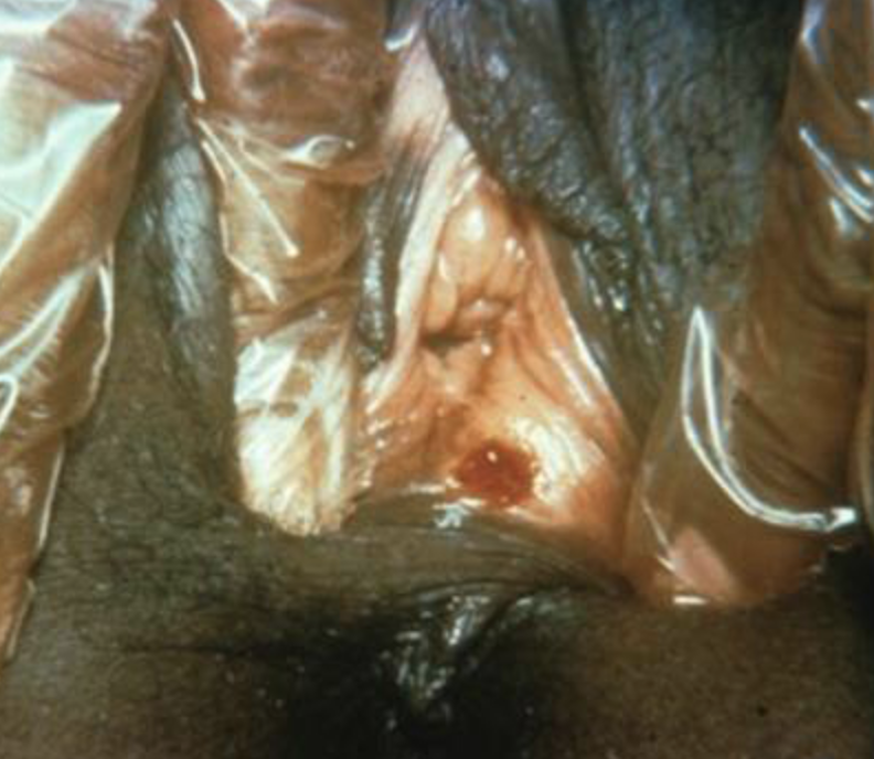
Most common presentation is a papule followed by a chancre or sore on the mouth, genitals or rectum
1 to 2cm with a raised margin
Appears on average 3 weeks after exposure with range is 10-90 days
Sore is painless and lasts 2 to 6 weeks but can be painful if co-infection present
Can be associated with bilateral lymphadenopathy, palpable on exam
Many do not notice the chancre which leads to increased transmission rates due to unknown or missed diagnoses
Frequent co-infection, test for other STIs
Reactive RPR, VDRL, FTA-ABS, MHA-TP, TP-PA, CIA, or EIA
Most common sign is diffuse macular, papular, or annular rash on the trunk and/or extremities | Pathognomonic is rash on palms of hands and soles of feet
Other symptoms: Ulcers in the mouth and genitals | Raised condyloma lata lesions near the primary outbreak
Systemic signs include weight loss, fever, malaise, alopecia and myalgia due to immune response to dissemination
Lymphadenopathy may be palpable in the femoral, inguinal, axillary and cervical regions
Other complications include uveitis, meningitis, nephritis, hepatitis, and synovitis
Reactive VDRL, RPR, FTA-ABS, MHA-TP, TP-PA, CIA, or EIA
Secondary stage syphilis sores (lesions) on the palms of the hands. Referred to as “palmar lesions”
Latent Syphilis
Seroreactivity with no symptoms
Develops 1 year to 30 years after initial infection and divided into early versus latent syphilis
Early latent syphilis: Infection occurred within the past 12 months
Late latent syphilis: Infection occurred more than 12 months ago, can last for years
If unknown duration of disease, classify as late latent
Cardiovascular and aortic involvement can occur, as well as tabes dorsalis (slow degeneration of the nerve fibers in the dorsal column of the spinal cord)
Reactive VDRL, RPR, FTA-ABS, MHA-TP, TP-PA, CIA, or EIA
Tertiary Syphilis
15 to 30% of untreated primary syphilis will develop tertiary
Develops 10 to 30 years after infection and can be fatal
Can cause blindness, deafness, destruction bone, mental illness, cardiovascular involvement, granulomatous lesions
Reactive VDRL, RPR, FTA-ABS, MHA-TP, TP-PA, CIA, or EIA
Neurosyphilis
Often divided into early and late neurosyphilis | Early characterized by meningitis, uveitis, or retinitis
Patients exhibiting tertiary syphilis should have CSF analysis, ophthalmic exam, and otologic exam
Symptoms of neurosyphilis can include: Abnormal gait | Lower extremity numbness | Difficulty in concentration or confusion | Headache | Seizures | Stiff neck | Depression or anxiety
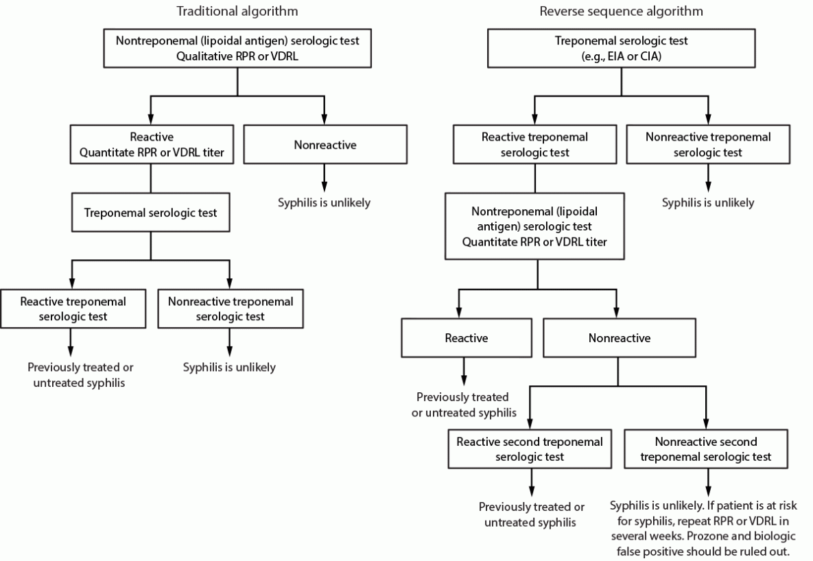
SCREENING TESTS AND ALGORITHMS:
Screening Tests
Screening for Syphilis Infection Is Always a 2-Step Process | There Is No Single Test For Syphilis
Treponemal test (assays detect IgM and IgG antibodies specific to T pallidum) | Cannot detect recent infection from past treated disease | Sensitivity between 97 to 100% | False positive as high as 40 to 80% (why reflex testing is necessary)
Microhemagglutination test for antibodies to T. pallidum (MHA-TP)
T. pallidum particle agglutination assay (TPPA)
T. pallidum enzyme immunoassay (TP-EIA)
Chemiluminescence immunoassay (CIA)
Nontreponemal tests: Can identify recent infection versus past disease
Rapid Plasma Reagin (RPR)
Venereal Disease Research Laboratory (VDRL)
Toluidine Red Unheated Serum Test (TRUST)
If a treponemal test is used for antepartum syphilis screening, all positive tests should be reflexed to a quantitative nontreponemal test (RPR or VDRL) to determine if past infection or active infection and needs treatment
If the nontreponemal test is positive, then a treponemal test ordered to confirm exposure to syphilis and not cross-reactivity or false positive
Obtain a final nontreponemal titer at delivery for all women diagnosed with congenital syphilis confirming maternal treatment | Allows for comparison to neonatal titer
Nontreponemal tests are not interchangeable when used to determine antibody titers; testing on follow-up samples must be performed with the same type of test
The T. pallidum particle agglutination test is the preferred manual treponemal test
Courtesy of CDC Figure 3
Treatment:
Penicillin G Is the Only Known Effective Antimicrobial for Preventing Maternal Transmission to the Fetus and Treating Fetal Infection
Dose for Primary, Secondary and Early Latent Syphilis (98% success rate)
Benzathine penicillin G 2.4 million units IM in a single dose (CDC), usually split 1.2 million units in each buttock
A second dose of benzathine penicillin 2.4 million units IM can be administered 1 week after the initial dose | Some practitioners routinely administer 2 doses in pregnancy over consecutive weeks with no more than 10d between injections
Dose for late latent syphilis or latent syphilis of unknown duration
Benzathine penicillin G 2.4 million units IM weekly for 3 weeks for a total of 7.2 million units
Dose for neurosyphilis
Aqueous crystalline penicillin G IV 18 to 24 million units per day, administered as 3 to 4 million units IV every 4 hours or continuous infusion for 10 to 14 days
Side effects
Jarisch-Herxheimer reaction occurs in 44% of pregnant women and is an acute febrile reaction frequently accompanied by headache, myalgia, fever, and other symptoms that can occur within the first 24 hours after the initiation of any therapy for syphilis
If treating after ≥24 weeks, consider treatment on L&D with continuous fetal monitor for at least 24 hours
Treatment Success
4x decline in titer, e.g., from 1:16 to 1:4 | Sero-reversion, or the loss of antibodies over time
Special Considerations for Treatment
Women without a history of treatment should be staged and treated accordingly with a recommended penicillin regimen
Pregnant women who miss any dose must repeat the full course of therapy
Women with a history of adequately treated syphilis who do not have ongoing risk do not require further treatment
If after treatment patient has persistently low nontreponemal titer (< 1:8) and clinical improvement, no additional treatment needed
Necessary to inquire about partner treatment
All women who have syphilis should be offered testing for HIV infection
Penicillin Allergy
Pregnant women with syphilis in any stage who report PCN allergy should be desensitized and treated with PCN
Neither tetracycline or doxycycline can be used in pregnancy
Data insufficient to support use of ceftriaxone and neonatal outcomes
Treatment Failure (or reinfection): Signs or symptoms that persist | 4x increase non-treponemal titer persisting for >2 wks
Reasons for treatment failure include: High bacterial load at the time of treatment and short interval between treatment and delivery
15%–20% of persons with primary and secondary syphilis treated with recommended therapy will not achieve the 4x decline in nontreponemal titer used to define response at 1 year after treatment
Retreatment: Weekly injections of benzathine penicillin G 2.4 million units IM weekly for 3 weeks is recommended
Post-Treatment Titers
Check titers at time of treatment as baseline
Ideally use same test and same lab for repeat titers
4x increase in titer after treatment is definition of treatment failure
A decline in titers does not mean a declined in the risk for congenital syphilis
Check titers monthly for those at high risk for reinfection or patients in geography with high incidence
Fetal Infection and Antenatal Surveillance:
When syphilis is diagnosed in the second trimester, perform ultrasound evaluation on fetus
Routine amniocentesis to confirm fetal infection is not recommended
Delivery and Postpartum:
Deliver for fetal indications including evidence of fetal hydrops, abnormalities of electronic fetal heart rate monitoring
Pediatrics present at delivery
Send placenta to pathology | Often large, pale and hydropic with chronic vilitis
Document maternal serologic status and have plan for follow up | Ensure reported to public health
PRIMARY SOURCES:
Sykes et al. Public Health Rep, 2021
Assessment of an outbreak in congenital syphilis (CS) in AZ looking at missed opportunities for diagnosis and cost of broad screening
Between 2017 and 2018 cases of CS quadrupled from 14 to 61 | Study period of January 2017 to June 2018 to assess for missed opportunities for diagnosis
Of 57 cases of CS in that period, 17 could have been prevented with third trimester screening
9 had prenatal care but screened late | 7 were infected after the first trimester screen | 1 was reinfected
Adding third trimester screening for all women in this system was balanced by the savings in treating CS in infants and saved $527 a year (AZ public insurance system analysis)
Conclusion
Third trimester screening for syphilis is cost effective and should be done in regions with high transmission
PROFESSIONAL RECOMMENDATIONS:
The USPTF recommends universal screening in pregnancy, and notes the recommendations of others in the final publication
This recommendation statement is consistent with those of other professional and public health organizations. The CDC recommends screening for syphilis infection in all pregnant women at their first prenatal visit
Joint guidelines from AAP and ACOG recommend screening for syphilis infection in pregnant women as early as possible in pregnancy
The CDC, AAP, and ACOG also recommend repeat screening at 28 weeks of gestation and again at delivery in high-risk women
Women at high risk for syphilis infection include those living in high-prevalence communities, those living with HIV, and those with a history of incarceration or commercial sex work
Defined as “Failure to deliver the fetal shoulder(s) with gentle downward traction on the fetal head, requiring additional obstetric maneuvers to effect delivery” (ACOG PB 178)
Shoulder dystocia is an obstetrical emergency
While there are associated risk factors, they are poor at predicting shoulder dystocia
The majority of cases will occur in women without diabetes whose offspring are within normal weight range | Nor is there any evidence that shoulder dystocia can be prevented
Complications include PPH and brachial plexus injuries | Severe neonatal morbidity can occur if shoulder dystocia is not resolved in a timely manner
Simulation of maneuvers been shown to improve use of maneuvers as well as teamwork and documentation
BACKGROUND:
Risk Factors
Maternal
Prior history of shoulder dystocia
Universal prophylactic cesarean section is not recommended
Due to recurrence risk (1% to 16.7%), evaluate EFW, GA, glucose and severity of previous event
Patient discussion and careful delivery planning are recommended
Diabetes: GDM and pre-gestational diabetes
Fetal
Macrosomia (see delivery recommendations below)
Large fetal chest relative to biparietal diameter (seen with diabetes)
Note: Despite known risk factors “…shoulder dystocia cannot be accurately predicted or prevented” (ACOG PB 178)
Evaluation
There are no ultrasound findings or labor patterns that are predictive of shoulder dystocia
The classic ‘turtle sign’ at delivery is “…suggestive, but not diagnostic, of the presence of shoulder dystocia” (ACOG PB 178)
Diagnosis is based on clinical judgement when there is failure to deliver the fetal shoulders after initial traction attempts
Maneuvers
See videos in ‘References’ below
McRoberts maneuver: Best first step (Level B Evidence)
Maternal knees flexed and brought to chest while suprapubic pressure is applied
Posterior Shoulder Delivery to reduce shoulder diameter (Level C Evidence)
Next option if McRoberts unsuccessful
Decreases the diameter of the fetal shoulder girdle
Additional techniques to deliver the posterior shoulder include the following
Rubin maneuver: Place hand on the back of the posterior fetal shoulder followed by anterior rotation towards the fetal face
Woods Screw maneuver: Apply pressure to anterior surface of the posterior shoulder with fetal rotation until anterior shoulder disengages from behind the maternal symphysis
Posterior axilla sling traction: Thread a size 12 or 14 French soft catheter around the posterior shoulder and apply moderate traction to the sling to deliver the shoulder
Gaskin all-fours maneuver (for women without anesthesia): With patient on hands and knees, apply gentle downward traction on the posterior shoulder or upward traction on the anterior shoulder
‘Last Resort’ maneuvers: Associated with significant maternal and/or fetal morbidity and mortality
Zavanelli maneuver: Head placed back in vaginal canal followed by cesarean section
Abdominal rescue: shoulder dislodged from above via hysterotomy
Intentional fetal clavicular fracture
Do not apply fundal pressure due to risk for uterine rupture
Instruct patient to stop pushing until dystocia is resolved
Evidence does not support use of routine episiotomy
Reserve for clinical situation where additional room may be needed for above maneuvers
Documentation should be contemporaneous and include (Level B Evidence)
Time of diagnosis
Management
Time of delivery
Sequelae
Simulation Programs
Simulation is used to train healthcare personnel for particularly severe, high acuity events that are relatively infrequent
Simulation is effective in the setting of shoulder dystocia and improves
Communication | Use of maneuvers | Documentation (Level B Evidence)
Management for Suspected Fetal Macrosomia
Delivery <39 weeks gestation is not recommended without medical indication
Elective cesarean delivery should be considered for the following
Without diabetes: estimated fetal weight of ≥5,000 gm
With diabetes: estimated fetal weight of ≥4,500 gm
Induction
Not suggested for suspected fetal macrosomia as induction has not been shown to improve maternal or fetal outcomes
Trial of labor
Suspected fetal macrosomia is not a contraindication to a trial of labor after cesarean delivery
PRIMARY SOURCES:
Zhang et al. BJOG, 2018
Meta-analysis to determine whether the maternal pre-pregnancy obesity has an impact on shoulder dystocia risk | 20 studies (2,153,898 participants)
Results
For obese versus nonobese: Pooled relative risk (RR) of shoulder dystocia was 1.63 (95% CI, 1.33–1.99)
Significant association remained across different continents
When adjusted for gestational diabetes the increased risk remained significant: RR 1.61 (95% CI, 1.05–2.47)
Compared to nonobese women, the pooled RRs for multiple obesity classes were as follows
Obesity class I: 1.29 (95% CI, 1.06–1.57)
Obesity class II: 1.94 (95% CI, 1.26–2.98)
Obesity class III: 2.47 (95% CI, 1.56–3.93)
Conclusion
Maternal pre-pregnancy obesity is associated with increased risk of shoulder dystocia
Higher the obesity class, the greater the risk
PROFESSIONAL RECOMMENDATIONS:
ACOG PB 178
The guideline states that Maneuvers may be repeated if not successful initially and in addition
…clinicians should use the maneuver most likely to result in successful delivery
No randomized controlled trials have compared maneuvers for shoulder dystocia alleviation
However, it is clear that brachial plexus injury can occur regardless of the procedures used to disimpact the shoulder(s) because all maneuvers can increase the degree of stretch on the brachial plexus
Second stage perineal pain relief in patients who have no regional anesthesia or have perineum sparing
Posterior perineum and vaginal repair if local anesthesia is insufficient
Posterior perineal pain after gynecologic surgery
Studies have shown failure of one or both sides in as many as 50% of transvaginal pudendal nerve blocks, assumed to be primarily due to failure of technique (Ford et al. J Obstet Gynaecol, 2013)
Generally considered safe when performed by a skilled clinician
Anesthetic effect
Some effect is seen after 5 minutes
Maximum effect at 10-20 minutes
Effect length: Varies depending on type of anesthetic used and half-life (approximately 30-60 minutes with lidocaine)
Lidocaine and 2-chloroprocaine are fast onset and short duration local agents | Bupivacaine and ropivacaine have slower onset but longer mechanism of action
Bupivacaine (longer acting agent) has a ‘black box’ warning for obstetric anesthesia
Reports of cardiac arrest and difficult resuscitation after using 0.75% concentration in epidural anesthesia
It is considered safe for use for pudendal block and included in the ACOG list of acceptable anesthetics
BACKGROUND:
Before widespread use of regional anesthesia for labor, the pudendal block was commonly used to alleviate the posterior perineal pain from the distention during the second stage of labor
Pudendal block does not provide relief for the following
Contraction or cervical dilation related pain
Relief to the anterior vulva or upper vagina
Known Complications
Hematoma
The pudendal nerve runs in the neural-vascular bundle under the sacroiliac ligament next to the ischial spine
Puncture of the vessels could lead to hematoma, a risk increased in patients with bleeding disorders
Neural injury
Rare, but lasting paresthesia can result (pudendal and sacral)
Infection
Localized infection can occur
Rarely involves spread to the hip joint, retro-psoas or gluteal space
Toxicity
If injected intravascularly, local anesthesia systemic toxicity can occur
Symptoms of systemic toxicity
Characteristic symptoms: Tinnitus and metallic taste in mouth
Severe complications: Seizures | Loss of consciousness | Arrhythmias | Respiratory and cardiac collapse
Procedure (see figure below)
Equipment: Pudendal Kit generally contains
Introducer
Spinal needle slightly longer than the introducer
Control syringe (there is often a small plastic spacer on the needle, remove this before using the kit)
20cc of local anesthetic
Epinephrine use (ACOG PB 209)
ACOG states that “Epinephrine may be added to local anesthetic solutions to delay absorption and increase duration of blockade by inducing vasoconstriction of the blood vessels in the area”
Has been associated in the past with increased need for oxytocin administration and vacuum assistance possibly due to loss of bearing down reflex in one study (Langhoff-Roos and Lindmark. Acta Obstet Gynecol Scand, 1985) | However, study results have not been corroborated
Can also serve as a marker for intravascular administration due to maternal tachycardia
Therefore, avoid in women for whom tachycardia is contraindicated
Injection technique
Using sterile gloves insert the left hand 2 fingers (with the introducer held between the index and third fingers) into the vagina and identify the left ischial spine
Approximately 1 cm medial and inferior to the spine is the sacrospinous ligament (which may be palpable)
Place introducer up against the ligament and advance spinal needle all the way though the introducer into the tissues, aspirate to ensure not in a vessel, then inject 10 cc of local anesthetic slowly
Repeat the procedure with an additional 10cc and the right hand on right ischial spine to obtain a bilateral block
Paracervical Block
Option to control uterine pain in labor, however, has fallen out of favor due to
Availability of more effective regional options
Need to repeat the procedure every 1 to 2 hours to maintain effect
Fetal bradycardia which can occur up to 10 minutes post block and can last up to 40 minutes
Incidence is estimated at 3.2% in a large Finnish database study (Palomäki et al. Acta Obstet Gynecol Scand. 2005)
Reports of this complication mostly note no long-term fetal impact though there are old reports of low Apgars and even fetal death (Shnider et al. AJOG, 1970)
PRIMARY SOURCES:
Novikova and Cluver. Cochrane Database of Systematic Reviews, 2012
Review of 12 RCTs of “unclear quality” | 1549 participants | Included both pudendal and paracervical nerve blocks in labor
Only 1 study compared nerve block (paracervical block) to placebo and 1 that compared paracervical nerve block to opioids
Both studies showed nerve block to be superior
Remaining studies looked at different agents compared to each other | Included both paracervical and pudendal blocks
Pace et al. Ann NY Acad Sci, 2004
RCT comparing single shot spinal anesthesia vs pudendal block in patients at >7 cm dilation
Spinal anesthesia was superior for pain relief during labor and delivery
Two approaches were statistically equivalent for pain control during perineal repair
PROFESSIONAL RECOMMENDATIONS:
ACOG PB 209
Pudendal block is useful primarily for the management of perineal pain in the second stage or to effect repair following delivery
Preterm birth: Defined as delivery between 20w0d and 36w6d gestation
Preterm labor: Defined by clinical criteria (ACOG PB 171)
Regular uterine contractions and documented cervical change (dilation, effacement, or both) or
Regular contractions and cervical dilation of ≥2 cm
In 2018, approximately 10% of all births in the U.S. were preterm which marks a continuous rise over the previous 4 years (March of Dimes Prematurity Profile)
Preterm birth accounts for 70% of neonatal deaths; 36% of infant deaths; 25-50% of long term neurologic impairment for children (ACOG PB 171)
< 10% of women with threatened preterm labor (preterm contractions) will give birth within 7 days of presentation | Increased rates related to shortened cervical length especially ≤15 mm (Fuchs et al. Ultrasound Obstet Gynecol, 2004)
Absolute risk of recurrent spontaneous preterm birth at <37 weeks’ gestation is 30% (Phillips et al. BMJ Open, 2017)
Risk of recurrence due to preterm premature rupture of membranes (PPROM) at <37 weeks: 7%
Risk of recurrence due to preterm labor at <37 weeks: 23%
Multiple gestation: Compared with singleton births, multiple births in the United States were about 8 times as likely to be preterm in 2017 (March of Dimes PeriStats)
Short cervical length: < 25mm before 24 weeks
Age: May be a ‘U’ shaped curve: 34-40 is associated with the lowest preterm birth rate | Odds ratio of 1.08 for women 20 to 24 years and 1.20 for women ≥40 years (Fuchs F, et al. PLoS One, 2018)
Race: The preterm birth rate is highest for black infants and lowest for Asian/Pacific Islanders (March of Dimes Peristats)
Black infants: 13.6%
American Indian/Alaska Natives: 11.3%
Hispanics: 9.4%
Whites: 9.0%
Asian/Pacific Islanders: 8.7%
Socioeconomic factors: Poverty is associated with preterm birth (Brumberg and Shah. J Neonatal Perinatal Med, 2015)
Previous surgical procedures for CIN (Kyrgiou et al. Cochrane Database of Systematic Reviews, 2017)
While excisional and ablative treatment increases risk for preterm birth, underlying CIN itself carries a baseline risk
Risk is higher with increasing cone depth and excision vs ablation
Low maternal BMI (<19.8)
Smoking and substance use
Interpregnancy interval: May be ‘U’ shaped curve with
increased risk < 18 months and >23 months | More recent data
indicate that risk range may be <12 months (Schummers et al. JAMA
Intern Med, 2018)
History and Physical
Review risk factors, clinical signs and symptoms
including
Back pain | Cramping | Pressure sensation in the
pelvis and/or vagina | Change in discharge or mucus | Bleeding or
spotting
Cervical exam
Increased dilation, effacement, softening or moving to
an anterior orientation | Speculum exam may demonstrate dilation on
visual exam, bulging membranes, bleeding or discharge to assess for
related complications such as PPROM, abruption or chorioamnionitis
Monitor with EFM
FHR: Assess fetal status and evidence of contractions
such as early decelerations
Tocometer: Presence and frequency of contractions
Imaging
If digital exam not advised (e.g., placenta previa)
Cervical length via ultrasound can provide additional
information | Cervical length is inversely related to risk for preterm
birth
Ultrasound may provide additional information
Can identify polyhydramnios, multiple gestation,
placental site location and fetal presentation
Laboratory Testing
Urinalysis and culture | CBC | GBS | Drug screen (if
index suspicion high for abruption) | Vaginitis or STI testing (as
indicated)
Role of Fetal Fibronectin (fFN) And Cervical Length
According to ACOG (PB 171), preterm labor is based on
clinical criteria and “the positive predictive value of a positive fetal
fibronectin test result or a short cervix alone is poor and should not be
used exclusively to direct management in the setting of acute symptoms”
Some centers have standardized protocols for preterm
labor that include the use of ultrasound for cervical length and
biomarkers such as fetal fibronectin (fFN)
SMFM states
fFN “may be more useful in managing symptomatic women with suspected preterm labor at/or before 34 weeks’ gestation” especially in women “with ‘borderline’ transvaginal cervical length measurements between 20 and 29 mm”
The combination of this intermediate cervical length
and fFN level >50 ng/ml may lead to consideration of corticosteroid
therapy for lung maturity
Recent meta-analyses suggest that use of ultrasound or
fFN may prolong pregnancy, however conclusive results will require further
research (Berghella and Saccone, Cochrane Database of Systematic Reviews,
2019)
Management <34 Weeks
Admission
Steroid administration
Tocolysis
Antibiotics for GBS prophylaxis
Magnesium sulfate for neuroprotection
ACOG/SMFM (Consensus Statement, 2017) recommend starting the above interventions as 24 weeks and should be ‘considered’ between 23w0d to 23w6d
Antenatal Corticosteroids
Corticosteroids reduce the following
Severity, frequency of respiratory distress syndrome | Intracranial hemorrhage | Necrotizing enterocolitis | Neonatal death
Steroid dosing
Betamethasone: (2 doses) IM 12 mg doses given 24 hours apart
Dexamethasone: (4 doses) IM 6 mg doses given 12 hours apart
Administer if there is risk of preterm delivery within 7 days
Between 24 to 34 Weeks Gestation
Intact Membranes: single course of corticosteroids
Ruptured membranes: single course of corticosteroids
Multiple gestation: single course of corticosteroids
Starting at 23 weeks gestation (irrespective of membrane status and fetal number): Single course of corticosteroids may be considered
Periviable period: Linked to family’s decision regarding resuscitation and should be considered in that context
Late preterm (34w0d to 36w6d) and No Previous Corticosteroids
Single course of betamethasone may be considered
Not indicated in women diagnosed with clinical chorioamnionitis
Note: Currently not known if late preterm corticosteroid
administration is of benefit in the setting of multiple gestation,
pregestational diabetes or previous course of corticosteroids
Rescue Dose
Regularly scheduled repeat courses or serial (> 2) courses are not recommended
May be considered in women who are
< 34 weeks gestation
Received a course of corticosteroids >14 days prior (can be lowered to within 7 days based on clinical scenario)
Are at risk of birth within the next 7 days
Whether to readminister steroids in women who are PPROM is controversial and there is insufficient evidence for or against
Magnesium for Neuroprotection (24 to 32 weeks)
Before 32 weeks, magnesium sulfate reduces the severity and risk of cerebral palsy
Women at high risk of delivery in the next 24 hours are candidates for magnesium for neuroprotection
Dose: 4 gm load over 20 min followed by 1 gm/hr for max 24 hours
Tocolysis
Goal is to safely administer antenatal corticosteroids
and complete steroid window for maximum neonatal effect
ACOG and SMFM
Consider 24w0d to be the lower age limit but consider
23w0d in specific clinical conditions
Recommend 34w0d as upper age limit for tocolysis
Contraindications to tocolysis in the setting of
preterm labor
IUFD | Lethal anomalies | Maternal bleeding |
Preeclampsia with severe features or eclampsia | Chorioamnionitis |
Abruption or other disorders where ongoing pregnancy is a greater risk
than early delivery
1st line tocolysis
ACOG: Beta-adrenergic agonist therapy, calcium channel
blockers or NSAIDs as first line, with option of magnesium sulfate
Other professional bodies, such as WHO and NICE
(guideline 25), recommend nifedipine as first line and do not recommend
magnesium sulfate or beta adrenergic receptor agonists
Dosing is inconsistent between studies and protocols
and may differ between centers (Haas. BMJ, 2012)
Maximum course of therapy: Up to 48 hours to allow for
the administration of antenatal steroids
Indomethacin
An NSAID that inhibits cyclooxygenase (enzyme that converts arachidonic acid to prostaglandin)
Administered up to 32 weeks
Not administered >32 weeks due to constriction of the ductus arteriosus
Side effects
Maternal: Nausea | Gastritis | Emesis
Fetal: Constriction of the ductus arteriosus | Oligohydramnios | Necrotizing enterocolitis
Dose: 50 mg load dose po | Maintenance 25 to 50mg every 6 hours | Important not to exceed 48 hours secondary to potential fetal side effects such as necrotizing enterocolitis (Hammers et al. AJOG, 2015)
Calcium channel blockers
(Nifedipine)
Block movement of calcium ions through calcium channels in cell membranes
Women with heart failure or reduced ejection fraction associated with preload abnormalities (e.g., aortic insufficiency)
Dose (most common dosing used in studies): 10 to 30 mg immediate-release nifedipine | Followed by 10 to 20 mg orally every 4 to 8 hours | Studies have reported various dosing regimens (Conde-Agudelo et al. AJOG, 2011) | No benefit to maintenance tocolysis
Preexisting tachycardia | Careful monitoring required in women with diabetes
Stop drug if pulse >120 bpm
Dose: 0.25 mg subcutaneously every 4 hours| IV not typically used (Haas et al. Int J Womens Health, 2014) | Other protocols include multiple doses initially
Note: FDA issued a black box warning (2011) regarding prolonged
use of terbutaline beyond 48 to 72 hours | Oral route is not effective and
contraindicated in pregnancy
Magnesium Sulfate
ACOG and SMFM consider magnesium sulfate as a tocolytic option for 48 hours to allow for steroid administration | Other professional bodies and authors recommend against use for this indication based on evidence showing limited effectiveness (NICE Guideline 25; WHO; Haas et al. Int J Womens Health, 2014)
Side effects
Maternal: Flushing | Nausea | Diaphoresis
Requires close monitoring for loss of tendon reflexes, respiratory and depression
Fetal: Potential risk for neonatal depression (dependent on magnesium levels)
Contraindications
Myasthenia gravis | Monitor closely in women with impaired renal function
Dose: 4 to 6 gm load IV over 20 to 30 min | Maintenance infusion 1 to 2 gm/hr
Toxicity: If signs of cardiac or respiratory collapse, administer calcium gluconate 10 mL of 10% calcium gluconate IV (1g total) over 3 min (i.e., slowly) to avoid hypotension and/or bradycardia
Combination with other medications
ACOG states that if magnesium sulfate is being used for neuroprotection and patient is still contracting, calcium channel blockers and adrenergic receptor agonists should be used with caution due to increased risk of side effects | Prior to 32 weeks, indomethacin is an option
Antibiotics
With intact membranes there is no indication for
antibiotic administration to improve maternal or neonatal outcomes | A
Cochrane review of 14 studies (7837 women) demonstrated antibiotics did
not improve neonatal outcomes and administration was associated with
increased neonatal deaths and CP (Flenady et al. Cochrane Database of
Systematic Reviews, 2013)
Alternative Therapies
Bedrest, hydration and sedation in asymptomatic women
at increased risk of preterm delivery have not been shown to be effective
at preventing preterm delivery
In addition, there are risks to bedrest including VTE
and bone loss, as well as socioeconomic and psychological consequences
Multiple Gestation
There is an increased risk of pulmonary edema with the
use of tocolytics in multiple gestations
There is neonatal benefit from antenatal corticosteroid
administration, thus most experts recommend steroids and tocolysis in for
women at risk of preterm labor with multiple gestations as well as
magnesium for neuroprotection
Progesterone and Prevention of Preterm Birth
Previous Spontaneous PTB (Singleton)
Risk assessment
Detailed medical history and prior obstetric history
Management
Insufficient data to recommend IM 17-OHPC
Serial endovaginal cervical length measurements starting at 16w0d and repeated every 1 to 4 weeks until 24w0d
If cervical length ≤25 mm, consider the following
Vaginal progesterone (vs cerclage)
Cervical cerclage (vs vaginal progesterone if not already on supplementation)
Physical exam indicated cerclage
Cervical pessary: Not indicated
No Previous History of PTB
Low risk for PTB
Clinical utility of universal cervical length screening “remains unsettled”
Cervix should be visualized at the 2nd trimester anatomy exam (18 to 22 weeks) | Transabdominal or endovaginal approach is acceptable
If cervix appears short on transabdominal scan, endovaginal ultrasound is recommended to determine whether progesterone may be of benefit
Serial endovaginal ultrasonography is not indicated in low risk patients
Short cervical length (≤25 mm)
IM 17-OHPC: Not indicated
Vaginal Progesterone: Indicated | “Although most studies used 200 mg progesterone daily from the time of identification of a cervix shorter than 25 mm at 18 0/7–25 6/7 weeks of gestation until 36–37 weeks of gestation, there are no adequate dosing studies or comparative trials, and there are insufficient data to indicate which formulation and which dose are most effective”
Cervical cerclage
Ultrasound-indicated: Overall, no significant reduction of PTB | May be potential benefit in very short cervix (<10 mm)
Physical exam-indicated: Consider if dilated cervix on digital/speculum exam at 16w0d to 23w6d “are candidates” for cerclage | Uncertain if amniocentesis to test for infection impacts outcome
Pessary: Not recommended
Multiple gestation with or without history of PTB
Cervix should be visualized at the 2nd trimester anatomy exam (18 to 22 weeks)
IM 17-OHPC: Not indicated
Vaginal progesterone: Insufficient data
Cerclage if cervix ≤25 mm
Ultrasound: Insufficient data
Physical exam-indicated: Consider procedure
Pessary is not recommended
History of a Medically Indicated Preterm Delivery
May be increased risk for PTB
“insufficient evidence to support a recommendation that these individuals undergo serial cervical length surveillance in future pregnancies”
PRIMARY SOURCES:
Crowther et al. Cochrane Database of Systematic Reviews, 2014
Meta-analysis of 37 trials (3571 women) that studied whether magnesium sulfate is an effective agent for the prevention of preterm birth
Magnesium sulfate was ineffective at delaying or preventing preterm birth when compared to no treatment, placebo or other tocolytics (e.g., betamimetics/calcium channel blockers/cox inhibitors, prostaglandin inhibitors)
Blackwell et al. Am J Perinatol,
2020 – The PROLONG Study
PROLONG study was a ‘confirmatory’ RCT performed with FDA input as a requirement for the accelerated approval pathway to determine whether IM 17-OHPC was effective for the prevention of preterm birth
A previous study, on behalf of the NICHD, demonstrated success of IM 17-OHPC in preventing preterm birth (Meis et al. NEJM, 2003)
Methods: Compared 17-OHPC IM injections to sham
Primary outcomes: PTB < 35 weeks and a neonatal morbidity composite index
Results: No differences seen in primary outcomes nor any of the individual components that were part of the composite index
Conclusion: “In this study population, 17-OHPC did not decrease recurrent PTB and was not associated with increased fetal/early infant death”
PROFESSIONAL RECOMMENDATIONS:
ACOG (PB 171)
A single course of corticosteroids is recommended for women between 24 to 34 weeks gestation at risk of delivery within 7 days | Consider single course starting at 23 weeks regardless of membrane status
Magnesium sulfate reduces the severity and risk of cerebral palsy in infants before 32 weeks of gestation
1st line tocolysis for up to 48 hours to allow for the administration of antenatal steroids is acceptable
Do not routinely prescribe antibiotics for pregnancy prolongation
Bed rest and hydration are not effective for prevention preterm birth and should not be routinely recommended
Do not use fFN or a short cervix alone to manage acute symptoms of preterm labor
Note: IM 17-OHPC is not recommended for the primary prevention of preterm birth in patients with a history of spontaneous preterm birth | Makena and related generics have been removed from the market
Most cases can be diagnosed based on history and physical examination
Avoid digital examination due to infection risk, unless delivery appears to be immediate
Membranes may reseal spontaneously leading to good outcomes
Hospital admission is recommended if the fetus is viable to monitor for signs of infection, abruption and fetal compromise
Acceptable strategy includes periodic ultrasound for fetal growth and FH monitoring (precise timing not established)
No clinical utility evidence for the use of serial WBC counts or other infectious markers
Use of tocolysis
Therapeutic tocolysis is not recommended
GBS as per standard protocol (see ‘Related Topics’ below)
GBS prophylaxis should be given based on prior culture results or intrapartum risk factors if cultures not performed or unavailable
If a patient is a candidate for prophylaxis, she should be given therapy “regardless of earlier antibiotic treatments” (ACOG PB 2017)
“If results are not already available and if an indication for treatment is not already present, culture for group B streptococci (GBS) should be obtained when expectant management is being considered.” (ACOG PB 217)
PROM-Related Risks
Preterm birth
Risks associate with prematurity include RDS, sepsis, IVH and NEC (van der Ham et al. PLoS Med, 2012)
Approximately 50% will delivery within the first week after membrane rupture (Lorthe et al. AJOG, 2018)
Latency period dependent on gestational age | Later gestational age more likely to deliver within a shorter timeframe (Walker et al. J Perinatol, 2014)
At term>60% begin labor spontaneously within 24 hours of rupture | >95% begin labor spontaneously within 72 hours (Hannah et al. NEJM, 1996)
Infection: (Kenyon et al. Cochrane Database of Systematic Reviews, 2013)
Preterm PROM and intrauterine inflammation are associated with increased risk of neurologic injury
Intraamniotic infection: may be up to approximately 30%
Postpartum infection: 5 to 20%
Maternal sepsis: approximately 1%
Abruption
Occur in approximately 5% of cases (Gonen et al. Obstet Gynecol, 1989)
Infection and umbilical cord accidents
Associated with a 1 to 2% chance for fetal demise
BACKGROUND:
Diagnostic Techniques
Speculum examination
Visualization of amniotic fluid (AF) leaking through the cervix
Vaginal pooling
Fern test of dried vaginal fluid seen under microscope
pH testing
Normal: 3.8 to 4.5
Amniotic Fluid: 7.1 to 7.3
False positives: Blood or semen, alkaline antiseptics or BV
False negatives: Minimal remaining AF following rupture
If above inconclusive
Ultrasound for AFV may be helpful but not diagnostic
Amniotic protein tests have high sensitivity for PROM but false-positive rates may be as high as 19% to 30%
ACOG (PB 217) states that “At most these test kits should be considered selectively relative to standard methods of diagnosis” (see FDA warning below)
Conclusive Test – Dye Instillation
Indigo Carmine
Ultrasound guided indigo carmine dye (amnio instillation) may be used, with passage into the vagina and detected with tampon or pad stain approximately 30 minutes later
Maternal urine may turn blue following instillation of indigo carmine
Fluorescein
2 to 5 ml 10% (200 mg to 500 mg) | 1 to 4 ml 5% (50 mg to 200 mg) has been suggested as sufficient (Ireland et al. Obstet Gynecol, 2017)
While there are potential IV side effects such as yellowing of sclera/palms, nausea, emesis and allergic reaction, no adverse events have been reported with amnio instillation
Original technique describes speculum exam of cervix at 15 and 45 minutes post injection using a long-wave ultraviolet light
Yellow-green fluorescent fluid leaking from cervix confirms the diagnosis
Fluorescence will rapidly appear in urine and confusion may be resolved with either visualization of cervical leak or tampon
Clinical Considerations for PROM at <24 weeks
Prospective EPIPAGE-II study (Lorthe et al. AJOG, 2018)
Gestational age at which PROM occurs impacts risk of mortality and cerebral palsy, with low survival at 22 and 23 weeks, but 70% survival at 24 and 25 weeks
Combination of birthweight, gestational age and sex will impact morbidity/mortality
Pulmonary hypoplasia
Will occur in approximately 10% to 20% of cases
Insufficient data to recommend ultrasound for determination of lung volumes or function
Oligohydramnios can result in Potter’s deformation sequence
ACOG advises that patients should be counseled regarding risks associated with expectant management vs intervention: “The rates of composite maternal morbidity (60.2% versus 33%) and severe maternal morbidity (12% versus 5%) were also significantly higher in the expectantly managed group” (ACOG Practice Advisory)
Management
Counsel regarding risks and benefits of expectant management vs immediate delivery
Immediate delivery should be offered as an option
Consider MFM and neonatology consultation
If patient chooses expectant management and no infection
Outpatient surveillance is an option
Give information to return to hospital immediately if signs or symptoms of bleeding, labor or infection (self-monitor temperature)
Advise return to hospital at time of viability
Corticosteroids and latency antibiotics
Data currently limited at <24 weeks
Offering antibiotics as early as 20w0d is an option
Consider offering a single course of corticosteroids as early as 23w0d of gestation due to risk of delivery within 7 days
Antenatal corticosteroids and latency antibiotics are recommended upon reaching viability
Preterm PROM at 24w0d – 33w6d
Expectant management in a hospital setting is recommended
If there are maternal and/or fetal contraindications to expectant management, delivery is recommended
There is evidence that delivery at 35 weeks may decrease NICU stay, although increase risk for short term infectious morbidities (Lynch et al. Am J Perinatol, 2019)
Antenatal (single course) corticosteroids are recommended
IV ampicillin (2 gm every 6 hours) and erythromycin (250 mg every 6 hours) for 48 hours followed by oral amoxicillin (250 mg every 8 hours) and erythromycin base (333 mg every 8 hours) for an additional 5 days (7 days total)
Azithromycin has been studied as an alternative to erythromycin | One such study finding no difference between standard regimen and patients who received azithromycin 1000 mg po once along with ampicillin IV (2 gm every 6 hours) for 2 days followed by amoxicillin po (250 mg every 8 hours) for 5 days (Navathe et al. AJOG, 2019)
ACOG (PB 217) states that azithromycin (e.g., 1 gm single dose) “is a suitable alternative” to replace erythromycin if unavailable or poorly tolerated
Amoxicillin–clavulanic acid
Not recommended due to increased risk for necrotizing enterocolitis
Allergy to β-lactam antibiotics
“May be reasonable to consider another agent against GBS” | Choice based on severity of allergic reaction and susceptibility profiling
Unclear as to whether cerclage should be removed or retained but if retained, antibiotic therapy should not be extended beyond 7 days
Patients with PROM from 24 to 32 weeks and at risk for imminent delivery within 24 hours are candidates for fetal neuroprotective treatment with magnesium sulfate if no contraindications (See “Related Topics’ below)
Dose: 4 gm load over 20 min followed by 1 gm/hr for max 24 hours
Obtain vaginal/rectal swab for GBS | Administer GBS prophylaxis as indicated
Late Preterm (34w0d to 36w6d)
“Either expectant management or immediate delivery is a reasonable option” (ACOG PB 217)
Data suggests when comparing these 2 options
No difference in neonatal sepsis
Newborn risks: Increased respiratory distress, mechanical ventilation, ICU stay in the immediate group
Maternal risks: Increased hemorrhage and infection in expectant management group
Administer single-course corticosteroids if
Not previously given
Delivery expected in >24 hours and ≤7 days
No chorioamnionitis
Screen for GBS and administer prophylaxis as indicated
Chorioamnionitis: Treat and plan for delivery
Term (≥37w0d)
Induction is recommended vs expectant management | Short period of expectant management (12 to 14 hours) “may be appropriately offered”
If no spontaneous labor, induce labor with oxytocin
Allow adequate time (12 to 18 hours) for latent phase to progress before performing a cesarean section for failed induction of labor
Induction with prostaglandins equally as effective as oxytocin but may have higher rates of chorioamnionitis
Insufficient data to recommend for or against cervical ripening with mechanical methods such as a Foley balloon
Insufficient evidence to recommend antibiotic prophylaxis beyond GBS indications
If a patient declines delivery and requests expectant management, counsel regarding risks and benefits
If fetal and maternal status are reassuring, expectant management ‘may be acceptable’
Screen for GBS and administer prophylaxis as indicated
Chorioamnionitis: Treat and plan for delivery
PROM Following Amniocentesis (ACOG PB 217)
Risk of PROM following amniocentesis is 1%
Outpatient, expectant management
Monitor regularly with ultrasound and counsel patients to watch for signs of infection, bleeding and/or miscarriage
Contrary to spontaneous PROM, good outcomes have been reported
Perinatal survival rate is approximately 90%
PPROM: HSV or HIV
HSV infection and Active Lesions (ACOG PB 220)
Risk of vertical transmission (CDC 2021 STD HSV Treatment Guidelines)
If HSV acquired near the time of delivery: 40 to 50%
If recurrent or acquired HSV during first half of pregnancy: <1%
Term PROM: Cesarean delivery is recommended in the presence of active HSV lesions or prodromal symptoms (e.g., vulvar pain or burning)
However, in the setting of PPROM
Risks of prematurity should be weighed against risks of neonatal vertical transmission
There is no established preterm gestational age at which immediate delivery recommended in the setting of PPROM
If expectant management planned, begin antiviral treatment (CDC 2021 STD HSV Treatment Guidelines)
Primary or first episode: Acyclovir 400 mg orally three times a day for 7 to 10 days or valacyclovir 1 gm orally twice a day for 7 to 10 days (extend treatment if healing complete after 10 days of therapy)
Symptomatic recurrent episode: Acyclovir 400 mg orally three times a day for 5 days or acyclovir 800 mg orally twice a day for 5 days or valacyclovir 500 mg orally twice a day for 3 days or valacyclovir 1 gm orally once a day for 5 days
Corticosteroid use
Balance risk between risk of transmission and pulmonary immaturity
HIV infection
Optimal
management is uncertain due to concern of vertical transmission with
PROM
Management
should include a physician with expertise in the management of HIV in
pregnancy and standard HIV guidelines should be followed
Most
recent data suggest that vertical transmission risk my not be increased if
the patient is on highly active antiretroviral therapy with a low viral
load and has received antepartum and intrapartum zidovudine
Management
should be individualized
If
gestational age is early, but patient is on appropriate therapy with a
low viral load expectant management may be appropriate
Preterm PROM and Future Pregnancies
Increased risk of recurrent PROM and preterm birth
Offer progesterone supplementation starting at 16 to 24 weeks
Consider cervical length screening
Consider cerclage for women with the following
Current singleton pregnancy
Prior spontaneous preterm birth <34 weeks
Cervical length <25 mm prior to 24 weeks
PRIMARY SOURCES:
Walker et al. J Perinatol, 2014
Review of 239,808 non-anomalous, inborn, infants between 23 and 34 weeks
PPROM in 15.5% >24 hours before delivery
Latency period
<24 h: 84.5%
1 to 7 days: 10.5%
8 to 14 days: 2.7%
15 to 21 days: 1.0%
21 to 28 days 0.4%
>28 days: 0.8%
Lorthe et al. AJOG, 2017
National prospective cohort study of 803 women with PPROM at 24 to 32 weeks
Explores if tocolytic therapy following preterm PROM (PPROM) is associated with improved neonatal or obstetric outcomes. In a multi-center, prospective, population-based cohort study
Results: When adjusting for confounders, primarily bias due to indication for treatment, tocolysis was
Not associated with increase survival without severe morbidity | Odds ratio 1.01 (95% CI, 0.94 to 1.09)
Not associated with increased latency ≥48 hours | OR 1.03 (95% CI, 0.95-1.11)
Not associated with histological chorioamnionitis | OR 1.03 (95% CI, 0.92-1.17)
The choice of tocolytic medication (oxytocin receptor antagonists or calcium-channel blockers) had no impact on outcomes
Conclusion: There does not appear to be any clinical benefit for the use of tocolytic drugs in the setting of PPROM
PROFESSIONAL RECOMMENDATIONS:
ACOG Practice Bulletin 217
Use
of ‘prelabor’ is in keeping with reVITALize terminology and is defined as
the ‘spontaneous rupture of membranes that occurs before the onset of
labor’
The
following recommendation is based on consensus and expert opinion
The outpatient management of preterm PROM with a viable fetus has not been sufficiently studied to establish safety and, therefore, is not recommended
FDA (2018)
In
August 2018, the FDA sent a letter to health providers to be
aware of rupture of membranes (ROM) test limitations, due to reports of
adverse events
The FDA letter
states that “health care providers may be over-relying on ROM test results
when making critical patient management decisions, despite labeling
instructions warning against this practice.”
To
promote awareness and aid in the proper use of the ROM test,
the FDA letter states:
The following limitations are typically stated in ROM device labeling
– A negative result does not assure the absence of membrane rupture
– False negatives may result if the amniotic sac has resealed or the position of the fetus has obstructed the rupture
– The presence of blood, meconium, anti-fungal creams or suppositories, baby powder, baby oil, or the use of lubricant with a vaginal exam may interfere with the device
– The test may not be accurate if sample collection and testing occurs after the timeframe recommended by the manufacturer
– To help protect patients and reduce the chance of adverse events, ROM tests should be part of an overall clinical assessment, which may include physical examination of the patient and testing to detect leaking amniotic fluid
Following delivery, women often do not show signs or symptoms of hypovolemia until significant blood loss has occurred (25% or more of blood volume)
While laboratory values can be useful in determining clinical status, resuscitation response, and guide certain management decisions, their limitations within the setting of active hemorrhage must be kept in mind
Uterine atony is the single most common cause of PPH (70% to 80%)
There is lack of evidence to determine which specific uterotonics are superior (ACOG PB 183) and choice can be left to clinician’s discretion
The following medications have been found to be effective for uterine atony
Medication Table
NOTE: Contraindications include hypersensitivity to the specific medication
Tranexamic Acid (TXA)
Large RCT (Lancet, 2017) demonstrated death due to bleeding was reduced by approximately 30% in the treatment group if started within 3 hours
TXA prevents clot breakdown and can be initiated alongside uterotonics
Administer 1g in 10 ml (100 mg/ml) of TXA intravenously at a rate of 1 ml per min (i.e., over 10 min)
If bleeding continued after 30 min or stopped and restarted within 24 hours of the first dose, a second dose of 1g of TXA could be given
Contraindications: Avoid in women if there is a clear contraindication to antifibrinolytic therapy such as a thromboembolic event in pregnancy
Adverse events: “There is no evidence of adverse maternal or neonatal effects” (WHO Recommendations)
Surgical Management Strategies
Vaginal
Cervical laceration repair
D&C for retained products
Bakri or other balloon placement for tamponade
Vaginal packing for extensive laceration
Interventional radiology
If available, uterine artery embolization by interventional radiology
Data have shown successful pregnancies following this intervention
Abdominal
Compressive sutures (B-lynch and others)
Vascular ligation (uterine artery or O’Leary sutures, ligating the utero ovarian ligaments)
Abdominal packing
Hysterectomy
Massive Hemorrhage Protocol
In the setting of massive hemorrhage, treatment of presumed or proven coagulopathy must be initiated
Place a 2nd large bore (16 gauge or larger) needle
Notify the team
Bring medications to the patient
Initiate massive transfusion protocol
Apply fundal massage
Insert a Foley catheter
Recommended RBC:FFP:Plt replacement ratio
In the range of 1:1:1
Available products
Packed Red Blood Cells (PRBC): Approximate volume 350 ml, expected to increase the Hct by 3% (Hgb increase of 1)
Fresh Frozen Plasma (FFP): Contains all clotting factors as well as albumin and is effective for reversing coagulopathy as well as volume expansion (it is isotonic) | It contains virtually no RBC or Platelets | 1 unit of FFP has a fibrinogen concentration of 2 to 4 mg/ml
Cryoprecipitate: Generated by centrifuging 1 unit of FFP and freezing the factors in a volume of 10 to 15 ml | Contains 80 to 120u of factor VIII and 150 to 250 mg fibrinogen | Not blood type specific
Platelets: Generally pooled from whole blood donations and each unit has approximately 5.5×10¹⁰ platelets in 50 ml | Generally come in 5 to 6 packs for transfusion | Not blood type specific
BACKGROUND:
PPH Definition
Cumulative blood loss ≥1,000 ml or blood loss accompanied by signs or symptoms of hypovolemia within 24 hours after the birth process (includes intrapartum loss) regardless of route of delivery (ACOG reVITALize)
However, if more blood loss than expected is observed postpartum (>500 ml for a vaginal delivery or >1,000 ml in a cesarean delivery)
Quickly evaluate uterus (including tone), cervix, vagina, vulva, and perineum
Assess for coagulopathies, lacerations and hematomas, in addition to atony
Maternal mortality
Post-partum hemorrhage is the leading cause of mortality for women worldwide and has increased by >25% since the 1990s in the US primarily due to increased rates of uterine atony
Though the rate of mortality in the US from PPH has decreased steadily since the 1980’s it still accounts for up to 10% of maternal mortality and is responsible for increased rates of transfusion and peri-partum hysterectomy
Safety bundles for PPH shown to significantly improve outcomes
50% reduction of maternal mortality in California since the institution of the CA Maternal Quality Care Collaborative (CMQCC) safety bundles
Populations who refuse transfusion of blood products (ie., Jehovah’s Witnesses) should have a plan in place for management of bleeding with guidelines on what products they will accept as well as access to a cell saver device
Risk Factors for PPH
Major risk factors for PPH include but are not limited to
Previous uterine surgery
Abnormal placentation
Chorioamnionitis
Prolonged labor or induction
Multiple gestation
Uterine fibroids
Magnesium sulfate use
Previous history of PPH
Grand multiparity
Polyhydramnios
Macrosomia
Available Toolkits
The CMQCC provides an online OB Hemorrhage toolkit which contains management flow sheets, educational tools, simulation resources, and checklists all of which are freely available
This safety bundle has been adopted by several states and in studies has been shown to contribute to reduced maternal mortality
Additional resources are available through the ACOG District II Safe Motherhood Initiative (SMI)
SMI has worked with over 10,000 obstetric providers to develop the contents of the maternal safety bundle toolkit that includes PPH
PRIMARY SOURCES:
Oladapo et al. Cochrane Database of Systematic Reviews, 2020
Meta-analysis to assess the comparative effectiveness and safety of IV vs IM oxytocin administration for prophylactic management of the third stage of labor following vaginal birth
Results
7 trials | 7817 women
IV oxytocin was associated with a lower risk for the following (high‐certainty evidence)
PPH (≥500 mL): Average risk ratio (RR) 0.78 (95% CI, 0.66 to 0.92)
Blood transfusion: Average RR 0.44 (95% CI, 0.26 to 0.77)
IV oxytocin probably reduces the risk of PPH ≥1000 mL (moderate certainty evidence)
There was a reduction in the risk of PPH ≥ 1000 mL with IV oxytocin in all but one study | If that one small study removed (only 3% of total events), the treatment effect favored IV oxytocin with high-certainty evidence
hypotension or other adverse events between IM and IV administration (moderate-certainty evidence)
Conclusions
IV oxytocin is more effective at preventing PPH following vaginal delivery when compared to IM without any substantive difference in adverse events
WOMAN Study: Lancet, 2017
The landmark WOMAN RCT sought to determine whether early administration of TXA could be beneficial for the treatment of PPH, similar to what was found in studies done in trauma settings
Methods: Randomized double-blind trial of TXA vs placebo given during PPH
20,000 women from 193 hospitals in 21 countries
Original endpoint was prevention of hysterectomy; however, the decision to use TXA was being made at the time of decision for hysterectomy, so the outcome of interest was changed to prevention of death
Results
Death due to bleeding was significantly reduced in the TXA group vs placebo (1.5% v 1.9%, RR 0.81 (95% CI, 0.65 to 1.0) and the impact was noted to be primarily within a 3 hour postpartum window
The Cochrane review of TXA studies to treat PPH demonstrated that there was
Reduced mortality risk related to bleeding, regardless of mode of birth
No increase in risk for thromboembolic events
Sentilhes et al. NEJM, 2018
RCT that evaluated the use of TXA for PPH prophylaxis
4079 women randomized to receive TXA or placebo
In addition, all patients received prophylactic oxytocin
Results
TXA did not demonstrate effectiveness for PPH prophylaxis
PROFESSIONAL RECOMMENDATIONS:
ACOG Practice Bulletin 183
“Postpartum Hemorrhage” guidance addresses the epidemiology, prevention and management of PPH
This bulletin includes management guidelines and medication dosing, as well as descriptions of available surgical techniques
There is also a section for low resource settings
ACOG Committee Opinion 794
“Quantitative Blood Loss in Obstetric Hemorrhage” guidance points out that visual estimation of blood loss can result in both over and underestimation
While quantification of blood loss in an important part of evidence based hemorrhage bundles, clinical utility specific to the quantification approach remains unproven and more research is needed
Timeframe for continuing blood loss assessment
Evidence is insufficient to recommend a precise time frame for ongoing measurement
ACOG suggests “that ongoing blood loss assessment should continue as long as active bleeding is present, or as long as the patient is unstable after a blood loss of more than 1,000 mL, including the postpartum care setting”
New Approaches: Artificial intelligence platforms
Artificial intelligence-based algorithms that use colorimetric analyses of pictures to quantify blood loss in real-time appear promising
ACOG states that while there is some supportive data, “validation of these findings with additional research is needed”
WHO
Based on evidence review, WHO also supports the use of tranexamic acid with postpartum hemorrhage
Early use of intravenous tranexamic acid (within 3 hours of birth) in addition to standard care is recommended for women with clinically diagnosed postpartum haemorrhage following vaginal birth or caesarean section
Peripartum cardiomyopathy (PPCM) is defined as heart failure that may develop toward the end of pregnancy or months after delivery without an identifiable cause (European Society of Cardiology)
While prognosis has improved substantially over the past several years, women with peripartum cardiomyopathy are still at risk for adverse outcomes (Honigberg and Givertz. BMJ, 2019)
Most women (50% to 80%) will make a full recovery (LVEF >50%) within first 6 months
For prognostic purposes, an LVEF ≥30% usually means a full recovery of left ventricular function is likely, while LVEF <30% suggests a slow or incomplete recovery with respect to achieving full ventricular function
Black ancestry is associated with increased risk and reduced likelihood of recovery
Unclear how much of this difference in outcome is due to genetic factors, socioeconomic status and/or structural racism (Williams et al. Health Psychol, 2016)
Due to increased recognition and improved treatment, mortality has improved from 30% to 50% in 1970’s to 1.3% to 16% in 2000’s (Honigberg and Givertz. BJM, 2019; Codsie et al. Obstet Gynecol, 2018)
Early consultation with a cardiologist/ MFM is recommended to coordinate care
If symptomatic pulmonary or peripheral edema present
Treat with loop diuretic
If hemodynamics permit
Selective β1 receptor blocker: Metoprolol preferred to avoid uterine stimulation via β2 pathway
Avoid ACE inhibitors and angiotensin receptor blockers (ARBs) during pregnancy
These medications may be used postpartum depending on lactation safety profile (enalapril and captopril acceptable)
Digoxin may be used in pregnancy
Anticoagulation: Data limited regarding precise threshold
AHA: LVEF <30%
ECS: LVEF ≤35%
Cardioversion and defibrillation may be used in emergent settings (safe in pregnancy)
BACKGROUND:
Risk Factors
Maternal age ≥30 years | Black Race | Hypertension | Anemia | Substance misuse | Asthma | Autoimmune disease | Preeclampsia or eclampsia | Multiple gestation | Obesity | Thyroid dysfunction | Prolonged tocolysis
Pathogenesis remains unknown but appears to be underlying background susceptibility with second ‘hit’ (e.g. endocrine factors of pregnancy)
Note: Preeclampsia and eclampsia are associated with PPCM and may have shared pathophysiology
Note: Not elevated in normal pregnancy | May be elevated in severe preeclampsia
Echocardiography is the most useful diagnostic tool
<45% LVEF (diagnostic requirement)
Right ventricular dilation (in some cases)
Pulmonary hypertension (in some cases)
Atrial enlargement (in some cases)
Atrioventricular valvular regurgitation (in some cases)
Differential Diagnosis
Benign dyspnea of pregnancy
Normal CXR | Normal echocardiogram
Asthma
Indicated by pulmonary function tests and bronchodilator response | Wheezing
Pulmonary embolism
Sudden onset |Tachycardia | Chest pain | Unremarkable pulmonary exam | DVT on LE imaging or PE on CT chest angiogram
Amniotic fluid embolism
Sudden onset | Circulatory collapse (during or after labor) | Bleeding (from DIC) | Hypotension | Tachypnea | Crackles on exam
Preeclampsia
Hypertension | Proteinuria | Usually accompanied by neurologic symptoms (headache, dizziness) | Echocardiogram shows mildly decreased LVEF
Treatment with Bromocriptine therapy
Sympatholytic dopamine D2 agonist
Experimental only, based on evidence that prolactin may be involved in pathogenesis
Associated complications: Lactation and thromboembolic events | Do not use without concomitant anticoagulation
Consider on an individual basis in severe cases (LVEF <25%) pending larger trials
Avoid over-diuresis to maintain perfusion of the placenta
Close monitoring throughout pregnancy and 6 months postpartum with echocardiograms (clinical scenario may dictate alternate/ more frequent regimen)
Each trimester
Immediately after delivery
4 weeks postpartum
Timing of delivery (AHA recommendations)
Stable: Per obstetric indications
Unstable or maternal extremis: Prompt delivery
Breastfeeding
Some controversy but unless severe LVEF, benefits may outweigh risks
Subsequent Pregnancies
LVEF prior to next pregnancy is the strongest predictor of outcome
If LVEF <50%
50% risk of acute heart failure with worsening disease and increased mortality
Pregnancy contraindicated without recovery to normal LVEF: “In women with a history of PPCM and persistent LV dysfunction, subsequent pregnancy carries a significant mortality risk and is clearly contraindicated” (AHA)
Ensure contraception counseling prior to discharge
Women with normal function prior to subsequent pregnancy are still at increased risk (20%) of worsening cardiac function
PRIMARY SOURCES:
Codsi et al. Obstetrics & Gynecology, 2018
Examined subsequent pregnancies of women with peripartum cardiomyopathy
Retrospective cohort study
Participants: All pregnant patients with prior peripartum cardiomyopathy defined as
Development of cardiac failure in the last month of pregnancy or within 5 months of delivery
Absence of an identifiable cause of cardiac failure
Absence of recognizable structural heart disease before the last month of pregnancy, and
Left ventricular (LV) systolic dysfunction with an LV ejection fraction (LVEF) <45%
25 patients with prior peripartum cardiomyopathy
60% of index pregnancies had a hypertensive disorder of pregnancy
3 (12%) patients with gestational hypertension, nine (36%) with preeclampsia, and 3 (12%) with confirmed eclampsia
12.1% of patients had a hypertensive disorder of pregnancy in subsequent pregnancies
Subsequent pregnancy (43 pregnancies)
Median gestational age at delivery for all live births in subsequent pregnancies: 39w0d weeks (range 36w6d to 41w3d)
All subjects except one had recovered LV function (LVEF ≥50%) before the subsequent pregnancy
Miscarriages: 6 (14.0%)
Terminations: 4 (9.3%)
Live births: 33 (76.7%)
Relapse rates
Rate of peripartum cardiomyopathy relapse: 20.9%
Median LVEF nadir in patients with relapse: 43% (range 35% to 45%)
Decline to the LVEF level of index pregnancy: 0
Relapse recovery: All patients with relapse recovered LV function within a median of 1 month
Cardiac arrests or deaths: None observed
Conclusions
Authors suggest the results of this study are encouraging, although further data would be required to determine optimal management in more diverse populations
PROFESSIONAL RECOMMENDATIONS:
ACOG PB 212
Due to related maternal morbidity and mortality, ACOG stresses the importance of identifying women at risk for cardiac disease as well as making a timely diagnosis
ACOG considers the ability to distinguish between common pregnancy symptoms (e.g., shortness of breath or palpitations) and cardiac disease to be “A key area of competence and expertise for obstetric care providers”
Furthermore, the document recognizes the work done by the CMQCC to develop a toolkit to aid in the above and recommends its use (see CMQCC link in ‘References’ for toolkit algorithm)
Maternal mortality reviews indicate that most women who die from cardiovascular disease had either undiagnosed cardiovascular disease or new-onset cardiovascular disease of pregnancy, specifically peripartum cardiomyopathy
Therefore, all women should be assessed for cardiovascular disease in the antepartum and postpartum periods using the California Improving Health Care Response to Cardiovascular Disease in Pregnancy and Postpartum toolkit algorithm
Use of this algorithm could have identified individuals as high risk requiring further cardiac evaluation and referral in 88% of maternal deaths
Lacerations are characterized by the structures they divide
The anal sphincter can be torn even if the perineum is intact
53% to 79% of women will incur a laceration at vaginal delivery (ACOG PB 198)
Majority are 1st and 2nd degree lacerations
27% of women at their first vaginal delivery have occult (concealed) injuries
Anatomy
The external anal sphincter (EAS) is comprised of circular striated muscle under voluntary control
The internal anal sphincter provides 80% of the resting pressure of the anal canal
The entire anal sphincter complex is 4cm in length
3rd and 4th degree lacerations are referred to as Obstetric Anal Sphincter Injuries or OASIS
3rd degree laceration: 3.3% of all vaginal deliveries | Divided into 3 types and includes injury to the perineal body in addition to
3a: Tear through ≤50% of the external anal sphincter
3b: Tear through >50% of the external anal sphincter
3c: Tear through the entire external anal sphincter and part of the internal anal sphincter complex
4th degree laceration: 1.1% of all vaginal deliveries
A tear through the external anal sphincter and the internal anal sphincter complex and includes the anal mucosa
A 2008 review of 451 articles and reviews noted that a “true” incidence of anal sphincter injury at vaginal birth is closer to 11% (Dudding et al. Ann Surg, 2008)
Simulation training for OASIS repair has been shown to be helpful
Routine episiotomy
Not recommended as it increases the likelihood and incidence of OASIS (Hartmann et al. JAMA, 2005)
Multiple reviews have found no maternal benefit to routine episiotomy
One review demonstrated the potential for increased incidence of postpartum anal incontinence, although uncertain due to quality of evidence | OR 1.74; 95% CI, 1.28 to 2.38 (LaCross et al. J Midwifery Womens Health, 2015)
Another meta-analysis of 22 studies demonstrated a significant increase in risk for OASIS injury with the use of midline episiotomy | OR 3.82; 95% CI, 1.96-7.42 (Pergialiotis et al. Int J Gynaecol Obstet, 2014)
BACKGROUND:
1st degree laceration: Separation of the skin and subcutaneous tissue of the perineal body, vagina or both
Most clinicians will repair periclitoral, periurethral, and labial lacerations that distort anatomy or are bleeding
If superficial with no bleeding, these lacerations can be left unrepaired
2nd degree laceration: Separation of the muscle of the transverse perineal body and/or the bulbocavernosus muscle body on either or both sides of the vulva
Continuous rather than interrupted sutures are recommended for 2nd degree repair
Episiotomy (see ‘Related Topics’)
If
episiotomy is warranted, mediolateral is preferred over midline given decreased
risk of extension into 3rd and 4th degree laceration | Studies show women may
have more pain and dyspareunia associated with mediolateral (ACOG PB 198)
Mediolateral
Risk of OASIS decreases by 50% for each additional 6 degrees from midline
Most providers cut at 45 to 60 degree angle
When comparing mediolateral episiotomy to no episiotomy at 3 months, risks were increased for (Sartore et al. Obstet Gynecol, 2004)
Perineal Pain: OR 3.1; 95% CI, 1.2 to 8.0
Dyspareunia: OR 2.4; 95% CI, 1.1 to 5.5
Midline
Episiotomy (midline) increases the length of perineal lacerations by approximately 3 cm compared to no episiotomy
Though not an accepted universal metric for quality, some institutions have adopted a target episiotomy rate of <5% | In 2012 the US rate was 12% (Friedman et al. JAMA, 2015)
Indications for episiotomy
Expedited delivery of the fetus
Fetal Heart Rate tracing abnormalities
Inability to monitor the fetal heart rate tracing when fetal vertex at outlet
Perineal tissue limits ability to perform shoulder dystocia maneuvers
Risk Factors for OASIS Injuries
The following have been identified as risk factors for OASIS (Pergialiotis et al. Int J Gynaecol Obstet, 2014)
Forceps assisted delivery: OR 5.50; 95% CI, 3.17 to 9.55
Vacuum assisted delivery: OR 3.98; 95% CI, 2.6 to 6.09
Midline episiotomy: OR 3.82; 95% CI, 1.96 to 7.42
Primiparous: OR 3.24; 95% CI, 2.2 to 4.76
OP position: OR 3.09; 95% CI, 1.81 to 5.29
Increased fetal birth weight: Mean difference, 192.88 g; 95% CI, 139.80 to 245.96
Previous 4th degree laceration: OR 10.55; 95% CI, 10.29 to 10.81
Prevention Strategies to Limit OASIS Injury
Studies have analyzed perineal techniques during the second stage of
labor (Aasheim et al. Cochrane Database of Systematic
Reviews, 2017)
Perineal Massage
Increased incidence of intact perineum: RR 1.74; 95% CI, 1.11 to 2.73
Fewer 3rd or 4th degree tears: RR 0.49; 95% CI, 0.25 to 0.94
Perineal Support
No difference in incidence of intact perineum
Fewer 3rd or 4th degree tears: RR 0.68; 95% CI, 0.21 to 2.26, five studies | Due to very low‐quality evidence, data should be interpreted with caution
Warm Compresses
No difference in intact perineum
Fewer 3rd or 4th degree tears: RR 0.46; 95% CI, 0.27 to 0.79
Steps for Repair of 4th degree
Identify the apex and provide adequate anesthesia | Use adequate lighting | Digital rectal exam can assist with assessment
A single dose 2nd generation cephalosporin is reasonable (ACOG PB 198)
Repair Anal Mucosa
Delayed absorbable 4-0 or 3-0 polyglactin, poliglecaprone or chromic in a running subcuticular fashion
Some will close the rectal muscularis in a separate interrupted or running layer
Place index finger of non-dominant hand in the rectum during repair of the mucosa
Identify the internal anal sphincter – visible as a thickening of circular smooth muscle and bring together the IAS over the anal mucosa
Steps for Repair of 3rd Degree
Repair external Anal Sphincter by grasping the ends with an Allis clamp, bringing the edges together and approximate just past the midline
Make sure to grab and include in suture, the fascial capsule in addition to the muscle
One method is to reapproximate in 4 planes using figure-of-eight sutures
Posterior | Inferior | Superior | Anterior
There is absence of evidence favoring end-to-end or overlap of the EAS during repair
Mediolateral Repair
Reapproximate transverse perineal and bulbocavernosus muscles
Taking a larger bite with the suture on the lateral side than on the medial side of the tear to reapproximate equally
Note: See ‘Repair of Obstetric Perineal Lacerations (Leeman et al. AFP, 2003)’ and ‘Repair of Episiotomy and Obstetrical Perineal Lacerations’ (Schmidt and Fenner. AJOG, 2024) in ‘References’ below for images related to repair of episiotomy and lacerations
Checklist
Watch out for retained sponges during complex repair
Before/after counts with recording
Postpartum
25% of women with an OASIS will experience a breakdown in the first 6 weeks postpartum
20% will experience a wound infection
Fistulas may develop, have a high index of suspicion for poorly healing lacerations at postpartum follow up | 9% of rectovaginal fistulas are associated with obstetric trauma
Stool softener and oral laxatives should be part of inpatient and discharge medications
Monitor to ensure able to void prior to discharge
NSAIDs, icepacks, sitz baths, topical anesthetics are helpful | Limited opioids as well with knowledge that they do increase risk for constipation
Pelvic floor exercises | Biofeedback and pelvic floor physical therapy are all helpful and complimentary in the healing process
Subsequent Pregnancy
Increased risk of another OASIS in women with a history of OASIS is about 3%
67% – 90% of women undergo a subsequent vaginal delivery
Indications to recommend cesarean delivery after OASIS
Experience of anal incontinence after delivery
Wound infection or repeat laceration repair
Psychological trauma
PRIMARY SOURCES:
Hartmann et al. JAMA, 2005
Systematic review of 26 episiotomy studies from 1950 to 2004
Examined evidence available regarding maternal outcomes of routine vs restrictive use of episiotomy
Conclusion
Authors found no maternal benefit to routine episiotomy
Angioli et al. AJOG, 2000
Retrospective case review of perineal lacerations from 1989 to 1995
50,210 women who met inclusion criteria
Conclusion
Aside from episiotomy (use had declined over the time of this study), the authors identified other risk factors for severe perineal lacerations, including birth weight, maternal age and operative delivery
Pergialiotis et al. Int J Gynaecol Obstet, 2014
Meta-analysis of 22 studies consisting of 651,934 women
Conclusion
Episiotomy was a significant risk factor for severe lacerations
Other risk factors for severe lacerations included heavier infants and operative vaginal delivery
PROFESSIONAL RECOMMENDATIONS:
ACOG Practice Bulletin 198
Rates of occult injuries are high, thorough examination following a vaginal delivery is essential to detect defects requiring repair
Warm compresses applied prior to delivery while pushing may decrease risk of 3rd and 4th degree lacerations | Perineal massage may be helpful as well
End-to-end or overlap repair of external anal sphincter are both acceptable
“A single dose of antibiotic at the time of repair is reasonable in the setting of obstetric anal sphincter injuries, but further research is needed to determine whether severe perineal lacerations warrant routine postpartum antibiotics to prevent complications”
Absorbable synthetic suture is recommended over chromic for repair of OASIS
Reapproximation of the IAS and EAS is essential for anal continence
All women in pregnancy should be screened for opioid and other substance use disorders with history and validated screening tools
Screening should be universal
Universal toxicology screening is not recommended
For women with a known use disorder, stabilization with opioid agonist therapy is recommended
Supervised withdrawal is not recommended as it is associated with high relapse rates from 59 to 90% (ACOG/ASAM CO 711)
Sudden withdrawal can lead to
Preterm contractions and labor | Restlessness | Abdominal pain | Nausea and vomiting | Tremors | Suicidal ideation | Death in the setting of excessive emesis and diarrhea resulting in heart failure and cardiac arrest
Infants born to women with opioid use (agonist therapy or substance abuse) should be monitored for withdrawal (also known as neonatal abstinence syndrome [NAS])
Monitoring should ideally occur within a neonatal abstinence program
Women who are stable on agonist therapy and participating in the care of their child should be encouraged to breastfeed
If women relapse into use of illicit drugs: Suspend breastfeeding
Patients with active substance use are at higher risk for STIs due to lifestyle factors and other comorbidities such as mental illness or PTSD
Screen more frequently for these conditions
BACKGROUND:
There has been a nationwide dramatic escalation in opioid prescriptions in the US over the last decades
Part of the solution has been to call attention to responsible prescribing by physicians, including ObGyns’ role in prescribing during pregnancy and postpartum
Standardized prescribing tools can be hospital-based or EMR-based and can help guide safe prescribing practices
ERAS protocols for surgical pain management have shown benefit in reducing opioid prescriptions, and likely decreasing diversion as well (Bergstrom et al. Gynecol Onc, 2018; Macones et al. AJOG, 2019)
A recent research letter reported that perinatal overdose deaths increased in Ohio 10-fold in the 10-year period between 2009 and 2018 (Hall et al. Obstet Gynecol, 2020)
The number of opioid prescriptions given to pregnant women increased during 2009 to 2018
Parallel to increase in prescriptions and use, there was a dramatic growth in NAS, which increased from 1.5/1000 births in 1999 to 6/1000 in 2013 [ACOG/ASAM CO 711])
Opioid use disorder is well defined in DSM-V and screening tools are available to determine if a patient has a use disorder
Some validated tools include: 4Ps | NIDA quick screen | CRAFFT among others (ACOG/ASAM CO 711; AIM Screening Tools available in ‘References’ below)
The Institute of Medicine developed the acronym SBIRT to call attention to the need for Screening, Brief Intervention and Referral to Therapy (see SAMHSA SBIRT below in ‘References’)
If excess prescriptions are suspected, the prescription monitoring program for that state can be queried by any practitioner (see PDMP-TTAC in ‘References’ below)
Obstetric Complications
Obstetric complications from opioid use in pregnancy are multifactorial and can be attributed to withdrawal, concomitant drug use, as well as possible nutritional and psychological comorbidities, and may include
Abruption
Fetal death
Chorioamnionitis
Fetal Growth restriction
Preeclampsia
Premature labor and delivery
Placental insufficiency
Postpartum hemorrhage
Opioid use, in any trimester, is not associated birth defects
Withdrawal
Signs and symptoms of withdrawal should prompt evaluation for opioid abuse
Nausea and vomiting, diarrhea
Abdominal pain
Muscle pain
Diaphoresis
Hypertension
Restlessness
Dilated pupils, watery eyes
Anxiety, discontentedness
Tremors, yawning or goosebumps
Withdrawal starts after 4 to 12 hours with short acting opioids such as heroin, oxycodone, morphine, hydromorphone, fentanyl, with long-acting preparations such as MS-Contin, long-acting fentanyl and methadone withdrawal starts after approximately 24 to 36 hours
Treatment
Treatment in Pregnancy with Either Methadone or Buprenorphine
Both require a monitoring and treatment program, and special licensing to prescribe
Methadone: Opioid agonist with a long half-life
Once daily dosing | Must be administered in person at a treatment program
It has many years of safety data in pregnancy
Crosses placenta | Low concentrations in breast milk
Dosage changes are common as pregnancy progresses
Methadone doses in pregnancy are not related to severity of NAS | Efforts to reduce dosages are not recommended
Buprenorphine: Partial opioid agonist
Lower chance of overdose
Can be prescribed by a registered physician and can be self-administered
Generally, does not require dose adjustment in pregnancy
There is some evidence of safety | However there are no long-term studies available looking at childhood and developmental outcomes
It may be associated with less severe NAS (Minozzi et al. Cochrane Database Syst Rev, 2013)
If a patient wishes to seek help the national helpline can guide them to treatment options through the SAMHSA National Helpline (see ‘References’ below)
If a patient is in a recovery program with monitored methadone dosing
They can generally be returned to these programs upon discharge from the hospital with contact from the inpatient team
These programs often are not open for dosing on Sundays, and may require an extra day’s stay in hospital to provide the Sunday AM methadone dose before discharge
PRIMARY SOURCES:
Peeler et al. AJPH, 2020
Secondary analysis of a statewide quality improvement database of opioid-exposed deliveries from January 2017 to April 2019 | Assessed rates of medication for opioid use disorder (MOUD) and NAS (they term it neonatal opioid withdrawal syndrome [NOWS]) by race and ethnicity in MA
Results
89.3% of women with MOUD were non-Hispanic White women
Prenatal medication treatment rates were lower for Black and Hispanic vs White women
Non-Hispanic Black women: Adjusted odds ratio (AOR) 0.34 (95% CI, 0.18 to 0.66)
Hispanic women: AOR 0.43 (95% CI, 0.27 to 0.68)
There was no difference in rates and severity of NAS
Conclusions
While this study is retrospective and is derived from the data of only one state, it points out a significant inequity in access to proper care during pregnancy based on race and ethnicity
Jarlenski et al. Drug Alcohol Depend, 2020
Retrospective analysis of National Inpatient Sample database | 53.4 million deliveries in the US between 2003-2016 in women ≥18 years
Outcome
Presence of any of the 21 indicators of severe maternal mortality identified by the CDC | Secondary analysis excluded transfusion due to lack of information on indication and number of units transfused
Study Objective
Analyze outcomes considered “near misses” for maternal mortality
Results
In women with opioid use disorder, the rate of severe maternal morbidity was elevated
18 to 34 years: aOR 1.51 (95% CI, 1.41 to 1.61)
≥35 years: aOR: 1.17 (95% CI, 1.00 to 1.38)
Stimulant use disorders (amphetamine, cocaine) were associated with higher risks | Cannabis was not associated with increased risk
Conclusions
While this is a large dataset with inherent bias of inaccurate reporting and misclassification, it does indicate that women with substance use disorders are at higher risk of poor outcome
PROFESSIONAL RECOMMENDATIONS:
ACOG/ASAM Joint Committee Opinion 711
ACOG and ASAM specifically note in the main conclusions that screening for opioid use and use disorders should be routine and universal, noting
Screening based only on factors such as poor adherence to prenatal care or prior adverse pregnancy outcome can lead to missed cases and may add to stereotyping and stigma
The Academy of Breastfeeding Medicine Protocol 21
This document addresses breastfeeding in many substance abuse situations but notes specifically that the effects of methadone in breastfed infants and the transmission of methadone in breastmilk have been studied and it is considered safe
The Clinical Protocol states
Those who have SUD or use substances during pregnancy or the postpartum period should engage in multidisciplinary prenatal and postpartum substance use care Individuals who discontinue nonprescribed substance use by the delivery hospitalization can be supported in breastfeeding initiation with appropriate follow-up Breastfeeding should be avoided during the use of nonprescribed opioids
OBG Project CME requires a modern web browser (Internet Explorer 10+, Mozilla Firefox, Apple Safari, Google Chrome, Microsoft Edge). Certain educational activities may require additional software to view multimedia, presentation, or printable versions of their content. These activities will be marked as such and will provide links to the required software. That software may be: Adobe Flash, Apple QuickTime, Adobe Acrobat, Microsoft PowerPoint, Windows Media Player, or Real Networks Real One Player.
Disclosure of Unlabeled Use
This educational activity may contain discussion of published and/or investigational uses of agents that are not indicated by the FDA. The planners of this activity do not recommend the use of any agent outside of the labeled indications.
The opinions expressed in the educational activity are those of the faculty and do not necessarily represent the views of the planners. Please refer to the official prescribing information for each product for discussion of approved indications, contraindications, and warnings.
Disclaimer
Participants have an implied responsibility to use the newly acquired information to enhance patient outcomes and their own professional development. The information
presented in this activity is not meant to serve as a guideline for patient management. Any procedures, medications, or other courses of diagnosis or treatment discussed or suggested in this activity should not be used by clinicians without evaluation of their patient’s conditions and possible contraindications and/or dangers in use, review of any applicable manufacturer’s product information, and comparison with recommendations of other authorities.
Jointly provided by
NOT ENOUGH CME HOURS
It appears you don't have enough CME Hours to take this Post-Test. We no longer offer Hours.
Leaving ObG Website
You are now leaving the ObG website and on your way to PRIORITY at UCSF, an independent website. Therefore, we are not responsible for the content or availability of this site